 |
|
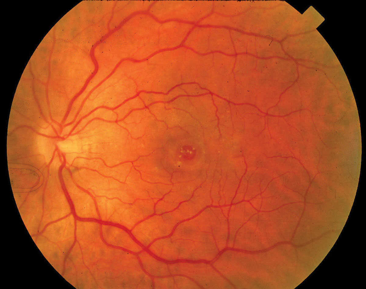
Idiopathic macular hole treatment with ILM peeling may have unfavorable visual outcomes. Photo: Diana Shechtman, OD. Click image to enlarge. |
Internal limiting membrane (ILM) peeling combined with pars plana vitrectomy and gas tamponade is an effective approach for treating traction maculopathies, but the findings of a recent retrospective case series suggest that though the procedure increases closure rate in these patients, it may also be detrimental to their retinal function.
The study included 45 eyes of patients with idiopathic macular hole who had undergone vitrectomy with ILM peeling with a six-month minimum follow-up. The researchers examined eyes using microperimetry MP-3 in the central 20° of the visual field and OCT-A in the central 6mmx6mm area.
The macular hole in each patient closed six months after surgery. The researchers observed significantly decreased retinal sensitivity in only the perifoveal temporal ETDRS sector (from mean 24.97 to 19.98). Additionally, 13% of patients developed 24 scotomas, 62.5% of which presented in the perifoveal temporal sector. The researchers also found bumps in the outer nuclear layer along with inner retinal dimples on B-scan images. These were mainly found in the perifoveal temporal sector (76.8%). The researchers noted that this finding hadn’t been reported previously in the literature. They also wrote that the incidence of outer nuclear layer bumps was significantly higher among patients with scotomas than those without (83% vs. 18%).
The researchers concluded that ILM peeling leads to functional changes in the retinas of patients with idiopathic macular hole, particularly in the perifoveal temporal macula. They believe that retinal layer distortion may underlie scotoma pathogenesis.
“Combining the anatomical and functional changes after ILM peeling, we hypothesize that damage to the Müller cells contributes to the pathogenesis of scotomas in the perifoveal temporal macula,” the researchers explained in their paper. “Müller cells are an integral component of the ILM, contributing to the formation of the b-wave on an electroretinogram. The recordings of focal macular electroretinogram demonstrated a limited and delayed recovery of the b-wave amplitude six months after surgery, possibly indicating dysfunction or physiological changes of the Müller cells.”
Tao J, Yang J, Wu Y, et al. Internal limiting membrane peeling distorts the retinal layers and induces scotoma formation in the perifoveal temporal macula. Retina. September 13, 2022. [Epub ahead of print]. |