 |
|
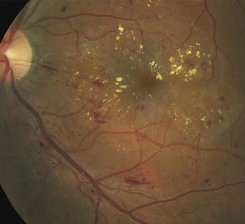
OCT-A determined that the total surface of capillary nonperfusion area was higher in eyes with DME than in eyes without DME in both superficial and deep capillary plexuses. Click image to enlarge. |
OCT-A for the first time is able to study both retinal vascular layers (superficial and deep capillary plexuses) and microstructure abnormalities without dye injection. However, little data is available on the role of microaneurysms, intraretinal microvascular abnormalities and capillary nonperfusion areas detected by OCT-A in diabetic macular edema (DME).
A recent study published in BMC Ophthalmology evaluated microaneurysms, intraretinal microvascular abnormalities, capillary nonperfusion areas, foveal avascular zone (FAZ) and capillary vessel density (VD) on 9mmx9mm OCT-A imaging in treatment-naïve DME eyes and compared it with findings in diabetic eyes without DME. It determined that this field of view was useful to detect retinal abnormalities beyond the 6mmx6mm field in order to have a more accurate analysis of the posterior pole. DME eyes showed a significantly higher number of microaneurysms, higher number of intraretinal microvascular abnormalities, larger FAZ area and larger surface of capillary nonperfusion areas on OCT-A than non-DME eyes. The capillary VD was significantly lower in DME eyes compared with non-DME eyes in the deep capillary plexus.
The researchers examined 70 eyes with diabetic retinopathy (37 eyes with DME and 33 eyes without DME). They reviewed and compared medical records, including swept-source OCT and 9mmx9mm swept-source OCT-A images between the two groups.
Compared with the non-DME eyes, DME eyes had more microaneurysms in the superficial and deep capillary plexuses, more intraretinal microvascular abnormalities in the superficial capillary plexus, larger areas of capillary nonperfusion in the superficial and deep capillary plexuses as well as a larger FAZ in both plexuses.
The capillary VD in the deep capillary plexus was lower in DME eyes compared with non-DME eyes. The severity of DME was significantly correlated to the number of microaneurysms and to the FAZ surface. Central macular thickness was significantly correlated with the number of microaneurysms in the deep capillary plexus, the surface of capillary nonperfusion areas and the FAZ area in both plexuses.
There was a significantly higher number of intraretinal microvascular abnormalities in DME eyes compared with non-DME eyes in the superficial but not in the deep capillary plexus. The researchers suggest this may be explained by the reduced visualization of vascular abnormalities in the deep capillary plexus due to the presence of intraretinal cysts.
Capillary vessel density is an objective tool on OCT-A to evaluate areas of nonperfused retina to improve DR monitoring and management. The researchers think the disorganization and nonperfusion of the deep capillary plexus play a major role in the occurrence of DME. Their results support the hypothesis that DME develops preferably in ischemic areas, which is consistent with the pathogenesis of DME.
“Our present findings could be useful to establish further objective and quantitative criteria for management and monitoring DME,” they concluded in their paper.
Braham IZ, Kaouel H, Boukari M, et al. Optical coherence tomography angiography analysis of microvascular abnormalities and vessel density in treatment-naïve eyes with diabetic macular edema. BMC Ophthalmol. November 3, 2022. [Epub ahead of print]. |