 |
|
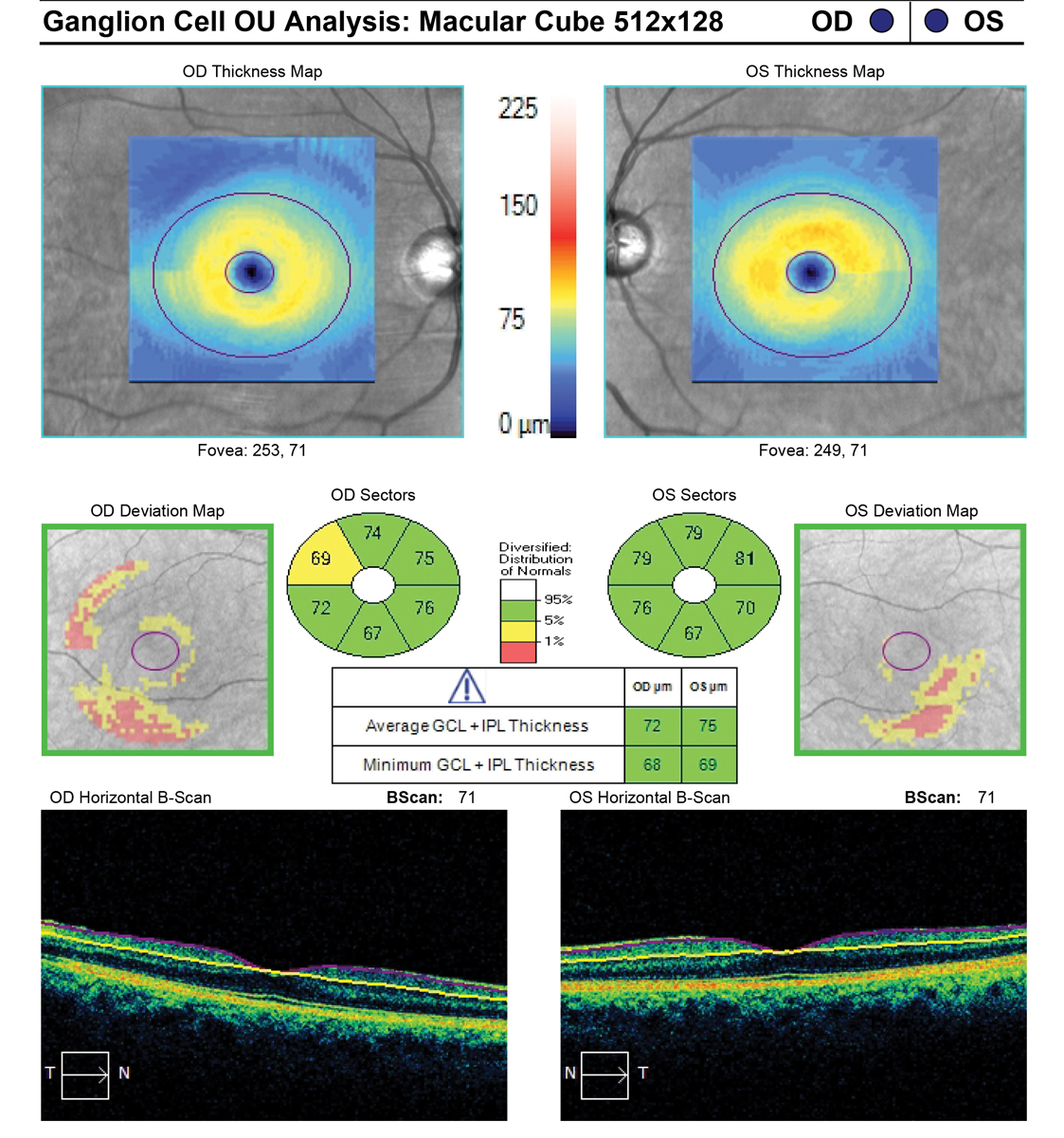
The full macular cube scan might prove more beneficial for diagnosing nonglaucomatous macular pathologies. Photo: Carl Zeiss Meditec. Click image to enlarge. |
The routinely used ganglion cell analysis (GCA) scan in OCT imaging is typically employed detect and monitor macular damage caused by glaucoma. While highly useful for that purpose, it can miss nonglaucomatous macular pathologies, new research finds. The study argues that the full macular cube scan might prove more beneficial in such cases.
The retrospective, cross-sectional study compared GCA printouts to full macular cube scans. GCA printouts provide information regarding the macular ganglion cell inner plexiform layer and a single B-scan of the retina, meanwhile the full macular cube scan includes all 128 B-scans. With a much higher amount of B-scans being done, the full macular cube scan was able to detect a fair amount of nonglaucomatous ocular pathology.
Included in the analysis were 105 patients and 201 eyes imaged with suspected glaucoma. In total, 32.3% revealed nonglaucomatous macular pathology through GCA printouts and macular cube scans. 38.5% of these eyes additionally included findings that did not show up on the GCA printout at all. Even further, 64.0% of those eyes that included macular pathology not visible upon printout required further evaluation.
While GCA is a convenient imaging test for rapid interpretation of qualitative and quantitative OCT data, a good amount of information outlining nonglaucomatous ocular pathology was unable to be seen, only to be discovered by the full series of B-scans offered in the full macular cube scan. All that is to say, the full macular scan might indeed provide more benefits in what it can detect.
The authors make note that the detection of some types of nonglaucomatous macular pathology, like ERM or IRF, are especially important, since changes like these that occur in the eye can actually end up confounding interpretations of the GCA. This occurs through segmentation altering if the GCIPL. As well, the nonglaucomatous macular pathologies found on these scans may turn out to need additional testing and treatment, thus making detection of utmost importance. As such, the full macular scan provides such preventative benefits through the better detection seen in this study.
The researchers believe that “these additional findings may alter clinical care decisions based on the discovery of nonglaucomatous macular pathology or by demonstrating artifacts that may influence interpretations of stability or progression. Clinicians should consider reviewing all the data that are captured to ensure they provide optimal patient care, although it is unknown if this missed pathology affects clinical outcomes at this time.”
Yamane M, Ferreyra H, Xu BY, Weinreb RN, Camp AS. Detection of nonglaucomatous macula findings with ganglion cell analysis printouts vs full macular cube scans. Sepetmber 8, 2022. JAMA Ophthalmol. [Epub ahead of print]. |