 |
|
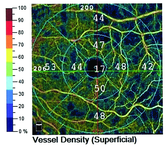
Macular microcirculation declined significantly in early POAG and NTG patients. Photo: Lori Mandy Pennington, OD, and Brooke Smith, OD. Click image to enlarge. |
Previous studies have reported decreased optic disc, peripapillary and macular vessel density in primary open-angle glaucoma (POAG) and normal-tension glaucoma (NTG) patients, showing comparable perfused 3x3mm macular and peripapillary capillaries between both groups but failing to establish the topographic difference in vessel and perfusion density. In a recent study, researchers compared macular vascular microcirculation in early POAG, NTG and controls and found that it declined significantly in affected patients.
A total of 99 patients with early glaucoma were included—60 POAG and 39 NTG. All underwent OCT-A scans at 6x6mm macular area. Macular vessel density and perfusion density of nine sectors were compared between groups. Linear regression analysis was used to investigate the relationship between vessel density and other variables including macular perfusion density, signal strength and mean macular ganglion cell-inner plexiform layer thickness.
Significant losses in total area of vessel density and perfusion density were detected in POAG and NTG compared with controls.
Similar to previous findings, the authors found that only the inner-inferior sector of macular vessel density decreased in NTG patients when compared with controls, while all inner sectors (superior, inferior, temporal and nasal) of macular vessel density were not significantly decreased in POAG. The macular perfusion density also followed the same trend between the NTG, POAG and control groups.
In early glaucoma, there were no differences in inner macular vascular parameters between the POAG and NTG groups.
The topographic differences of macular vessel density and perfusion density were obvious when compared with the controls, the authors noted. All sectors of the outer vessel density and perfusion density decreased in the NTG group, while only the superior-outer, inferior-outer and temporal-outer sectors of macular vessel density and perfusion density decreased in the POAG group.
The results of previous studies suggested that NTG is associated with earlier involvement of macular structural and functional defects. In this study, a more significant decrease in the inferior-inner and nasal-outer sectors of macular microvasculature was found in NTG patients compared with POAG patients.
“While the sample size of our NTG group analysis is limited, it appears that NTG patients have a more central and nasal macular microcirculation loss than in the POAG patients,” the authors explained in their paper. “Therefore, the topography of macular microcirculation seems to be a potential way to investigate the intraocular pressure-independent differences between NTG and POAG. However, there is still overlap between macular vessel density and perfusion density of eyes with POAG and eyes with NTG.”
Further studies on the macular microcirculation assessment are required to investigate the relationship between POAG and NTG, the authors concluded.
Huo Y, Thomas R, Guo Y, et al. Topographic differences in superficial macular vessel density in eyes with early primary open-angle glaucoma and normal tension glaucoma. Ophthalmic Res. January 5, 2023. [Epub ahead of print]. |