 |
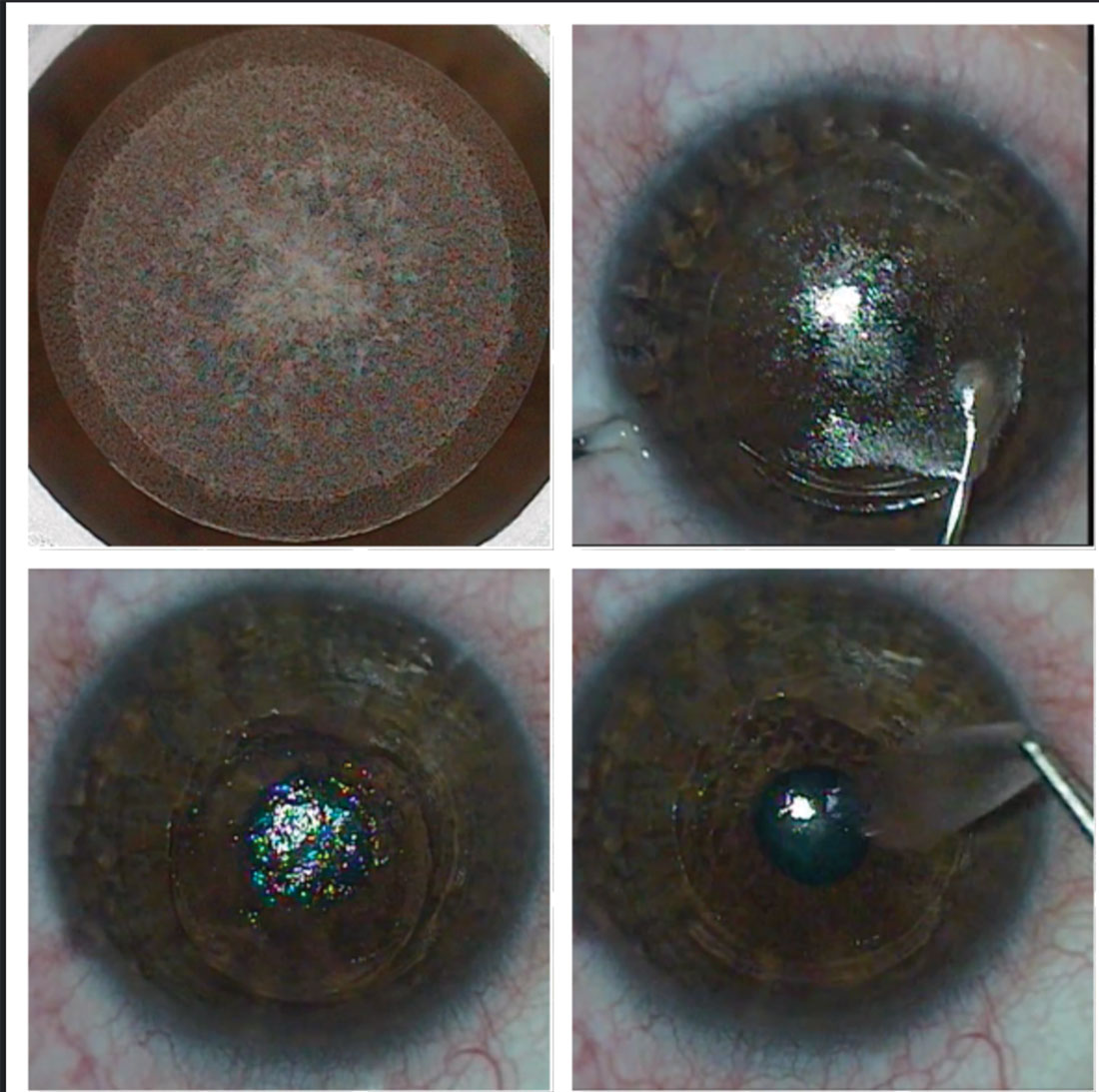
Patients who received SMILE demonstrated better image quality than those who received PRK or FS-LASIK. Photo: Bobby Saenz, OD, Anthony Vanrachack, OD, and Alexandra Wiechmann, OD. Click image to enlarge. |
When it comes to myopia correction, photorefractive keratectomy (PRK), femtosecond laser assisted in-situ keratomileusis (FS-LASIK) and ReLEx small incision lenticule extraction (SMILE) have all been found to be equally effective with minimal post-op issues. Stacking all three procedures against each other, a new study found SMILE had better image quality and similarity between eyes, in addition to providing closer pre-op values.
The study team from India suggests their results can be explained from the underlying increase in higher-order wavefront aberrations experienced by the eye after surgery.
“While the three keratorefractive procedures tested here are efficacious at correcting the spherocylindrical refractive errors of the eye, they all increase the magnitude of higher-order aberrations that adversely impact the patient’s postoperative optical quality and visual performance,” the authors wrote in their paper. “The results of this study clearly demonstrate that the extent of postoperative deterioration in image quality is not equal between procedures.”
Specifically, the study found that SMILE produced the least amount of increase in higher-order aberrations (1.6-times), followed by FS-LASIK (2.8-times) and PRK (3.3-times), relative to their preoperative values.
The study enrolled 106 participants, 40 each in the FS-LASIK and SMILE groups, in addition to 26 individuals who underwent PRK. The researchers considered wavefront aberrations and image quality of both eyes in all study participants preoperatively and again at one week, one month, three months and six months post-op using computational through-focus analysis for 6mm pupil diameter. Image quality was based on peak value and its interocular difference, residual defocus needed to achieve peak image quality (best focus) and depth of focus.
One month after surgery, higher-order aberrations were less frequent after SMILE, about 0.34μm compared with PRK (approximately 0.80μm) and FS-LASIK (roughly 0.74μm), relative to pre-op values (approximately 0.20μm).
Following PRK and FS-LASIK, peak image quality dropped, while interocular difference increased, best focus shifted myopically by 0.5D to 0.75D and depth of focus widened significantly. All these changes were negligible but still statistically significant in a minority of instances following SMILE surgery.
While previous studies showed spatial and depth vision estimates were better following SMILE, the current investigation provides the optical basis for these findings using image quality analysis, the authors explained.
Sarkar S, Devi P, Vaddavalli PK, et al. Differences in image quality following three laser keratorefractive procedures for myopia. Optom Vis Sci. December 29, 2021. [Epub ahead of print]. |