 |
|
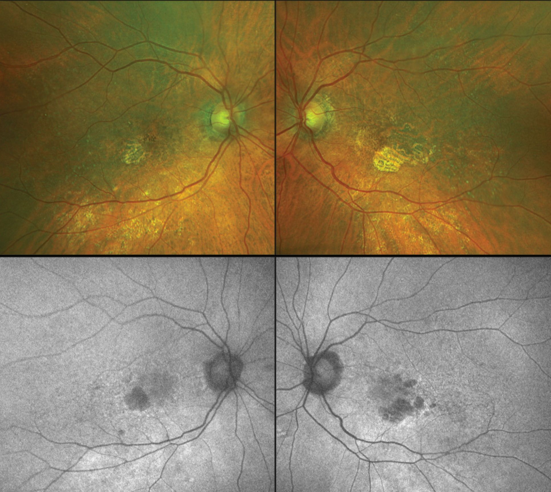
A new form of autofluorescence imaging was able to differentiate two potential disease pathways in geographic atrophy, originating from intermediate AMD lesions, to help simplify and aid research. Photo: Anna Bedwell, OD. Click image to enlarge. |
The pending release of new therapies expressly targeting geographic atrophy is bringing renewed focus to this aspect of age-related macular degeneration (AMD), as its mechanisms are complex and still not completely understood. Fundus autofluorescence is reliably used for identifying retinal pigment epithelium loss based on decreased autofluorescence, but there’s often residual autofluorescence from fluorophores below the retinal pigment epithelium, resulting in nonuniform autofluorescence levels that often appear dark or gray.
Recently, researchers proposed a way of addressing these gray areas through a new technique called quantitative autofluorescence (qAF), which uses “a fixed internal fluorescent reference to account for variable laser power and detector sensitivity,” according to a paper published by the group in the journal Eye (London). This approach allows for objective stratification of individual geographic atrophy lesions and also—when paired with serial SD-OCT use—reveals the origins and pathways to each geographic atrophy lesion in cases of intermediate AMD (iAMD). Using this technique, they were able to identify two potential distinct pathways of geographic atrophy, stemming from iAMD lesions.
In the study, the researchers imaged 23 eyes of 18 patients using SD-OCT and qAF. They selected 52 geographic atrophy regions of interest (i.e., clusters of adjacent lesions) and divided these into groups based on the dominant iAMD precursor from patients’ previous SD-OCT scans. Then, they compared the mean qAF values and structural SD-OCT findings.
Group 1 consisted of soft drusen/pigment epithelial detachment precursors (18 of 52 regions). The researchers reported that this group’s lesions were isolated and had a significantly lower mean qAF (35.88 units) vs. group 3, which consisted of multilobular subretinal drusenoid deposit precursors (12 of 52 regions) with a mean qAF of 71.62 units. The greater qAF demonstrated by the latter phenotype is likely due to the fluorescence of retained basal laminar deposits, according to the researchers. Group 2 included mixed precursors (22 of 52 regions), with an intermediate mean qAF of 58.13 units.
Based on these findings, the researchers reported that qAF can serve as a surrogate marker for differentiating geographic atrophy pathways originating from soft drusen and subretinal drusenoid deposits. They pointed out in their paper that by dividing the disease into two components, each could be studied separately, and this “might improve the interpretation of research on geographic atrophy mechanisms and prognostic factors for disease progression.”
Wei W, Mazzola M, Otero-Marquez O, et al. Two potentially distinct pathways to geographic atrophy in age-related macular degeneration characterized by quantitative fundus autofluorescence. Eye (Lond). January 9, 2023. [Epub ahead of print]. |