 |
|
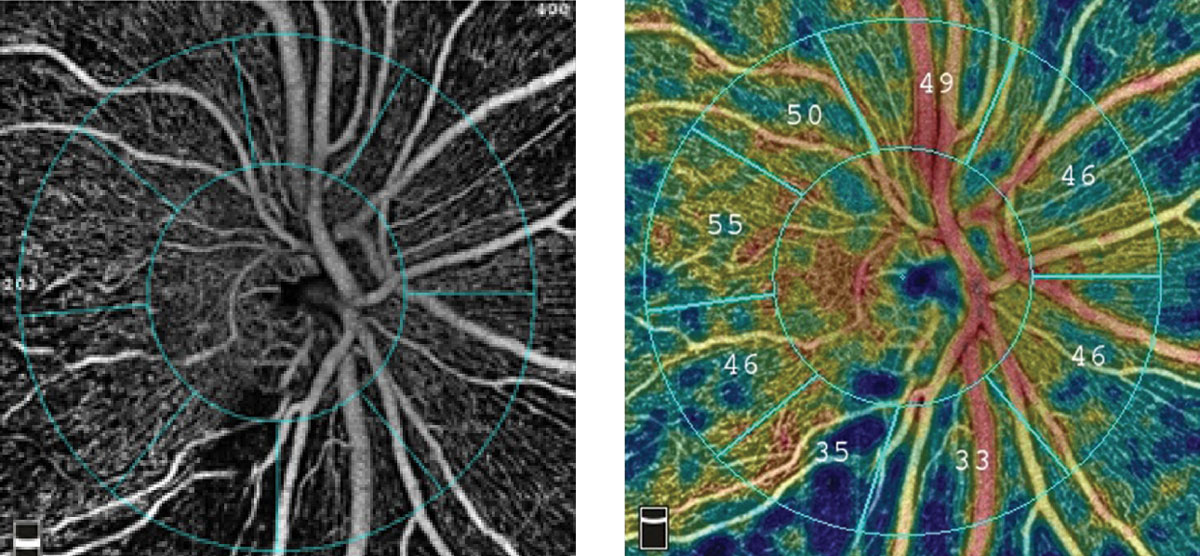
Glaucoma testing two to three times a year was found to be enough to detect progression in this study. Photo: Optovue. Click image to enlarge. |
Glaucoma’s all-but-inevitable advancement poses clinical and logistical challenges to eyecare providers when setting a follow-up schedule that isn’t onerous for patients but doesn’t run the risk of missing significant findings. A new study sought to determine how testing intervals affect how much time is required to determine further disease development. Progression was measured in two different ways. One included looking for the circumpapillary retinal nerve fiber layer (cpRNFL) with OCT and the other method looked at circumpapillary capillary density (cpCD) with OCT angiography (OCT-A).
The retrospective, longitudinal study included 156 total eyes and patients with glaucoma follow-up with a 3.5 year average. All participants had at least four OCT or OCT-A tests to better determine longitudinal rates of cpRNFL thickness and changes to cpCD.
What the researchers found was that an increased frequency in testing resulted in less time needed to map out changes indicative of progression on either measure. For eyes with cpCD loss of -1% per year, the follow-up time to identify at least 80% of cases dropped from six years when testing occurred once annually to four years when patients were tested three time per year; in those tested semi-annually, it took 4.23 years to identify progression, suggesting this may be the “sweet spot” for frequency.
Similarly, in eyes with cpRNFL loss of -1μm per year, 80% were detected after 6.3, 5.0 and 4.2 years at the same testing intervals, making cpCD a slightly quicker mechanism in detecting glaucoma progression.
Overall, results indicated that after two years and having either two or three tests per year, there might be slight improvement in the detection of glaucoma progression for those with a higher vessel loss rate. Subtle improvement in detection may occur after five years for those with comparatively lower vessel loss rates.
Half of the eyes were found to progress at -2% per year after two years, as measured by OCT-A with a frequency of two tests per year. A -0.5μm loss per year was found in cpCD and measured by OCT-A for 60% of eyes after five years.
When establishing a monitoring schedule for early glaucoma, the researchers found that two tests a year was sufficient using both cpRNFL and cpCD methods to detect progression and they suspect OCT-A may be able to detect early changes that occur due to the disease. Likewise, they point out that increasing the number of tests from two to three per year does not reduce how much time it takes to detect progression and that two tests work just fine.
The authors point out this testing frequency will not work for everyone, advising that “several factors should be considered when defining testing frequency. For example, patients with advanced glaucoma or who have mild damage at a younger age may require more frequent OCT/OCT-A testing. Clinicians should consider for decision making other factors such as those for risk of progression including ethnicity, age and life expectancy.” They also make sure to note that both types of testing used in this study are only “one component of known factors in clinical practice that can show the patterns of glaucomatous damage,” indicating that glaucomatous damage detection may indeed be shorter in practice.
Mahmoudinezhad G, Moghimi S, Proudfoot JA, et al. Effect of testing frequency on the time to detect glaucoma progression with OCT and OCT angiography. Am J Ophthalmol. 2022. [Epub ahead of print]. |