 |
|
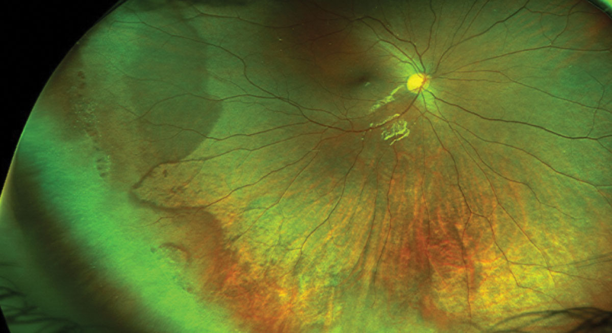
A highly myopic fundus may be best observed through ultra-widefield fundus imaging, which can capture portions of the eye that conventional imaging cannot. Photo: Mohammad Rafieetary, OD. Click image to enlarge. |
Posterior staphyloma is a hallmark sign of a highly myopic fundus and is associated with a significantly increased risk for progression of myopic maculopathy. Examining this portion of the eye through conventional fundus photography can be challenging due to its limited 50° view, especially for patients with a wide macular staphyloma (the most common type). Ultra-widefield fundus imaging covers up to 200° of the fundus, potentially making it a more ideal tool for identifying posterior staphyloma in highly myopic patients. Researchers recently put the two imaging modalities to the test, confirming the clear advantage of ultra-widefield over conventional photography in observing this condition and monitoring the progression of myopic maculopathy.
The study included 390 eyes of 210 highly myopic patients who completed follow-up for a minimum of five years using ultra-widefield fundus imaging, as well as various other imaging modalities, to monitor fundus abnormalities and signs of myopic maculopathy progression.
The analysis concluded that the extent of chorioretinal atrophic lesions and the presence of posterior staphyloma or peripheral retinal lesions is better documented through ultra-widefield fundus imaging than through conventional fundus photography due to the wider field of view. The ultra-widefield method was able to detect posterior staphyloma in 51% of eyes, while conventional fundus photography was not able to identify the border of staphyloma in 21% of those patients within its narrower field of view. In accordance with other studies, this analysis also showed a strong association between posterior staphyloma and the long-term progression of myopic maculopathy.
“During a mean follow-up period of 69.2 months, progression of myopic maculopathy was observed in 51.8% of eyes and was significantly associated with the presence of posterior staphyloma,” the researchers wrote. “The results of this study imply that identifying the presence of posterior staphyloma and the full extent of chorioretinal atrophy based on ultra-widefield fundus imaging may be clinically important in the follow-up of highly myopic eyes to monitor the pathologic alterations in the posterior pole.”
Considering most highly myopic patients with posterior staphyloma have the wide macular type, choosing ultra-widefield over conventional imaging may help to gain a clearer picture of a patient’s fundus changes over time and the status of myopia maculopathy progression.
Oh BL, Park UC, Kim BH, et al. Role of ultra-widefield imaging in the evaluation of long-term change of highly myopic fundus. Acta Ophthalmol. August 17, 2021. [Epub ahead of print]. |