 |  |
Neurotrophic keratitis (NK) is a degenerative disease of the cornea that leads to loss of corneal sensation. If left untreated, it can lead to perforation of the cornea and permanent vision loss.1-2
Patients with NK often present with a chief complaint of fluctuating vision, with little to no complaints of discomfort. They will have decreased corneal sensation in at least one quadrant of the cornea—this is the hallmark of the disease.3 Ocular surface findings could include persistent punctate epithelial keratitis, irregular/rough epithelium, neovascularization of the cornea, decreased tear prism, persistent epithelial defect, corneal ulceration, and—worst-case scenario—perforation.
There are several viable medical treatments for NK, and we will describe a surgical option when conservative treatments fail to regain corneal sensation: corneal neurotization.
Corneal Neurotization
This minimally invasive surgical procedure is performed under general anesthesia and grows new corneal nerves from a donor nerve tissue by either an autograft or allograft.4 When using an autograft or allograft, there are multiple ways a surgeon can re-establish sensory innervation to a neurotrophic cornea. Techniques for autograft use ipsilateral or contralateral supraorbital and/or supratrochlear nerves to reinnervate a neurotrophic cornea. Newer minimally invasive techniques use an acellular nerve allograft, which may minimize the risk of undesirable outcomes such as long visible scars, facial nerve injury and donor site morbidity.4
Here we will describe minimally invasive corneal neurotization using a nerve allograft.
Nerve Allograft
Eyelid crease incisions are performed to harvest the supraorbital or supratrochlear nerve. In some cases, the infraorbital nerve can be used. The surgeon will either use the supratrochlear or supraorbital nerve based on best-fit diameter to the nerve allograft.4
 |
|
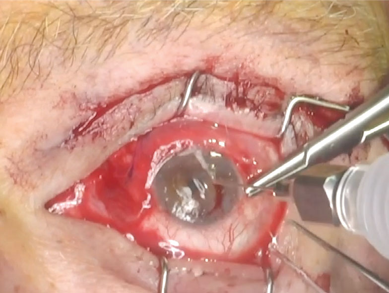
Nerve branches are secured to the sclera to help re-establish sensory innervation during corneal neurotization. Photo: Ilya M. Leyngold, MD. Click image to enlarge. |
For contralateral nerve transfer, bilateral eyelid crease incisions are made, and for ipsilateral transfer, unilateral incisions are made. Contralateral nerve transfer is done in cases where there is ipsilateral trigeminal neuropathy. After isolation of the donor nerve, it is then severed one to two centimeters below the incision site. The processed nerve allograft is then sutured to the donor nerve, and a nerve connector or amniotic membrane is used to protect the severed nerves and aid in the joining of the allograft to the host nerve.4
The nerve graft is tunneled through a blepharotomy incision to the contralateral side. Nerve fascicles are released from the graft through an incision in the epineurium and then tunneled to the corneoscleral limbus in the subconjunctival space. The perineurium of the nerve fascicles are then sutured to the sclera. Lastly, a permanent or temporary tarsorrhaphy is performed to protect the cornea while reinnervation takes place. The eye is then patched for a 24-hour period.4
Postoperatively, patients are instructed to use a topical antibiotic QID for one week. All preoperative drops that were being used to medically manage NK are resumed after removal of the eye patch. Patients are advised against strenuous activity or heavy lifting for two weeks.4
Dr. Besecker is founder of Envision Specialty EyeCare & Dry Eye Center in Meridian, Idaho. He has no financial disclosures.
1. Bonini S, Rama P, Olzi D, Lambiase A. Neurotrophic keratitis. Eye (Lond). 2003;17(8):989-95. 2. Labetoulle M, Baudouin C, Calonge M, et al. Role of corneal nerves in ocular surface homeostasis and disease. Acta Ophthalmol. 2019;97(2):137-45. 3. Dana R, Farid M, Gupta PK, et al. Expert consensus on the identification, diagnosis and treatment of neurotrophic keratopathy. BMC Ophthalmol. 2021;21(1):327. 4. Leyngold IM, Yen MT, Tian J, et al. Minimally invasive corneal neurotization with acellular nerve allograft: surgical technique and clinical outcomes. Ophthalmic Plast Reconstr Surg. 2019;35(2):133-40. |
