 |
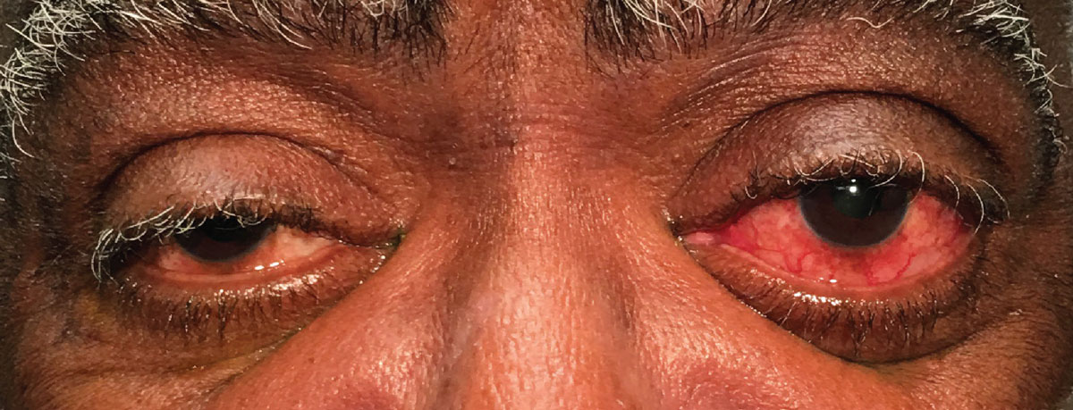
A 74-year-old man presented to the office with a chief complaint of a red and swollen eye of six days’ duration. He was pseudophakic OU. He reported some pain upon moving his eyes and mild tenderness upon palpation. He was not symptomatic for vision changes or diplopia. He denied any trauma. He was properly medicated for hypertension and diabetes and denied allergies of any kind.
Clinical Findings
His best-corrected entering visual acuities were 20/25 OD and 20/25 OS. His external examination is demonstrated in the photograph below. There was no evidence of afferent pupillary defect. His confrontation visual fields were full. His anterior segment examination revealed a normal OD and injected slightly proptotic OS. Goldmann applanation tonometry measured 17mm Hg OU.
Dilated fundus exam demonstrated no posterior pole or no peripheral pathologies with cup/disc ratios of 0.3/0.3, with sharp and pink margins.
 |
|
Our patient presented to the clinic with the presentation seen here. He reported no visual changes but some pain upon movement. Click image to enlarge. |
Other Testing
Additional studies included Hertel exophthalmometry, manual retropulsion of the left globe, binocular exam of the fundi to look for disc edema and choroidal folds, as well as neuroimaging, which included fat suppression MRI, venography and arteriography.
What’s the Culprit?
The diagnosis in this issue is orbital pseudotumor, or idiopathic orbital inflammation (IOI), involving the left medial rectus. Idiopathic orbital inflammatory diseases are generally defined as being noninfective, and are characterized by nonspecific inflammation within the orbit without identifiable local or systemic causes.1-8
IOI is typically benign and confined to the orbit.1-8 Once it forms it does have the capability to extend.1-4 Orbital involvement may progress into the sclera and/or nerve, producing periscleritis and perineuritis.1 IOI is often classified into categories of anterior, diffuse, posterior or apical, myositis and dacryoadenitis.1,10 The condition also has the capability to produce a focal orbital mass.1 Ocular myositis is a common form of IOI that frequently manifests with an acute and unilateral periorbital pain, swollen eyelids, conjunctival hyperemia, extraocular muscle movement impairment, proptosis and diplopia.8-10 Affected children often present with bilateral disease.10
IOI is not a disease per se; rather, it is a sign resulting from an immune system response.1-3 When it is discovered, testing must be completed in the attempt to expose the underlying disease.1-8 Differential diagnosis ranges from infectious disease (e.g., lyme disease, orbital cellulitis), autoimmune disease (e.g., thyroid eye disease, sarcoid, Crohn’s disease) to allergic, neoplastic/metastatic and idiopathic cases.3,4
The etiology of orbital myositis is unknown.5-7,9 The condition most commonly affects young adults in the third decade of life, with a female predilection.9,10 Whether acute or chronic, orbital myositis consists of localized inflammation, which occurs independently of the size of the affected muscles.7 Pathologically, orbital myositis has the potential for infiltration of inflammatory cells into degenerated extraocular muscles fibers.7 In these cases, inflammation of the extraocular muscles may lead to chronic ocular motility restriction in the non-acute phase secondary to fibrosis and destruction of extraocular muscle fibers.5-7 Orbital myositis may be part of a more widespread systemic inflammatory process such as Crohn’s disease or the recently described IgG4-related disease.8
Management Approach
When the clinical and radiological findings of an orbital inflammatory condition or mass are inconclusive, tissue biopsy—obtained by minimally invasive approach using local anesthesia—has been advocated.3,4 An orbital biopsy is usually done for accessible orbital lesions only.3 In cases of extraocular myositis and orbital apical inflammation, where surgery is difficult or dangerous, orbital biopsy is not initially considered.3
IOI is a diagnosis of exclusion.1-10 Some feel the diagnosis can be made according to clinical improvements after systemic steroid therapy; however, since other inflammatory and malignant disorders also response well to steroid therapy, histopathologic evaluation is still considered a mainstream of diagnosis.10,11
The mainstay of therapy for IOI extraocular muscle myositis consists of systemic corticosteroids.1-10 Other options include external beam radiotherapy, antimetabolites, alkylating agents, T-cell/calcineurin inhibitors, lymphocyte inhibitors, tumor necrosis factor-α inhibitors and surgical debulking.1
This patient was started on 80mg oral prednisone and responded well, with symptoms subsiding within four days and resolution occurring within 20 days. The medication was tapered slow over the course of two months. There was no residual loss of function.
Dr. Gurwood is a professor of clinical sciences at The Eye Institute of the Pennsylvania College of Optometry at Salus University. He is a co-chief of Primary Care Suite 3. He is attending medical staff in the department of ophthalmology at Albert Einstein Medical Center, Philadelphia. He has no financial interests to disclose.
|
1. Yeşiltaş YS, Gündüz AK. Idiopathic Orbital inflammation: review of literature and new advances. Middle East Afr J Ophthalmol. 2018;25(2):71-80. 2. Ding ZX, Lip G, Chong V. Idiopathic orbital pseudotumour. Clin Radiol. 2011;66(9):886-92. 3. Mombaerts I, Rose GE, Garrity JA. Orbital inflammation: Biopsy first. Surv Ophthalmol. 2016t;61(5):664-9. 4. Mombaerts I, Bilyk JR, Rose GE, et al. Consensus on diagnostic criteria of idiopathic orbital inflammation using a modified delphi dpproach. JAMA Ophthalmol. 2017;135(7):769-776. 5. Kinori M, Ko AC, Alameddine RM, et al. Extraocular muscle fibrosis in idiopathic orbital inflammation. J Pediatr Ophthalmol Strabismus. 2016;53(4):256. 6. Sousa L, Romano LM. Isolated medial rectus orbital myositis as a manifestation of idiopathic orbital inflammation. Acta Neurol Belg. 2011;111(1):78-9. 7. Kubota T, Kano H. Assessment of inflammation in idiopathic orbital myositis with fat-suppressed T2-weighted magnetic resonance imaging. Am J Ophthalmol. 2007;143(4):718-20. 8. Fraser CL, Skalicky SE, Gurbaxani A, McCluskey P. Ocular myositis. Curr Allergy Asthma Rep. 2013;13(3):315-21. 9. Costa RM, Dumitrascu OM, Gordon LK. Orbital myositis: diagnosis and management. Curr Allergy Asthma Rep. 2009;9(4):316-23. 10. Eshraghi B, Sonbolestan SA, Abtahi MA, Mirmohammadsadeghi A. Clinical characteristics, histopathology, and treatment outcomes in adult and pediatric patients with nonspecific orbital inflammation. J Curr Ophthalmol. 2019;31(3):327-334. 11. Swamy B.N., McCluskey P., Nemet A. Idiopathic orbital inflammatory syndrome: clinical features and treatment outcomes. Br J Ophthalmol. 2007;91(12):1667–1670. |
