 |
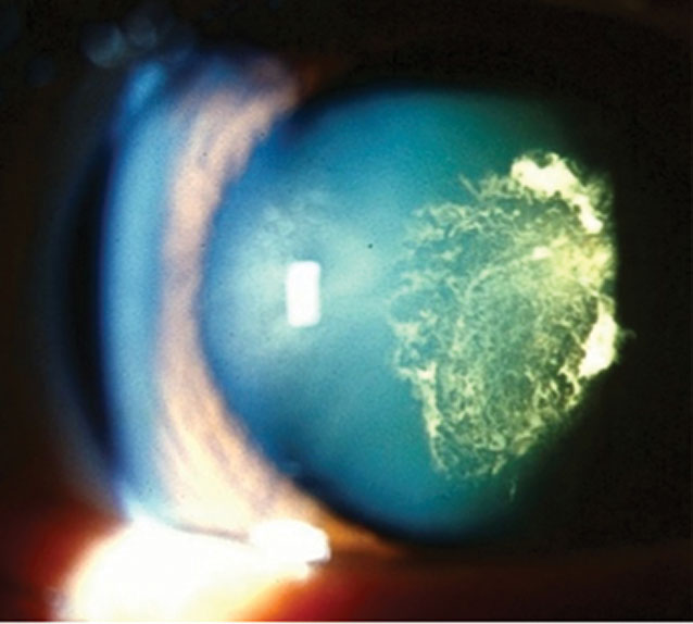
| The impaired detection of perfusion deficits within the central macula of cataract eyes needs to be appreciated when imaging the choriocapillaris in phakic eyes. Photo: Julie Tyler, OD. Click image to enlarge. |
As the sensitivity of ocular imaging technology increases, so does the potential for error. When the goal is to deliver ever more precise measurements, even small artifacts can affect the results. That seems to be the case when using OCT-A to image the fine blood vessels of the choriocapillaris behind a cataract, according to a small study conducted at Bascom Palmer.
Researchers investigated the impact of cataracts on image quality and the ability to measure macular choriocapillaris flow by comparing the quantitative results before and after cataract surgery using a novel OCT-A quality map algorithm and a validated strategy for quantifying the choriocapillaris. They found that the presence of cataracts corresponded with worse image quality and increased choriocapillaris flow deficits measurements within the central 1mm circle, which would suggest a decrease in the ability to accurately assess choriocapillaris perfusion in the presence of cataracts.
In 24 eyes, swept-source OCT-A image quality scores and macular choriocapillaris flow deficits measurements within the fovea-centered 1mm, 3mm and 5mm diameter circles were compared before and after cataract surgery. Choriocapillaris flow deficits changes in a modified ETDRS grid were further investigated.
Although overall image quality improved after cataract surgery, this improvement primarily impacted the measurements of choriocapillaris flow deficits in the 1mm fovea-centered circle, and there were no significant changes in the quadrants within the 3mm and 5mm circles.
Although the overall image quality of the study’s scans did improve after cataract surgery, there were seven cases in which the image quality decreased, and these decreases were primarily due to an increase in postsurgical vitreous opacities and a variability in the corneal tear film, as the use of artificial tears was at the discretion of the OCT operator.
The main limitations of the study were the relatively small sample size and the absence of a formal grading system to assess cataract severity, but the researchers wanted this study to reflect a real-world population of patients undergoing cataract surgery.
The researchers warned that, “Although the described technique should work for OCT-A scans with other fields of view, with different resolutions and from different manufacturers, it has not been validated previously for such use and, due to the machine learning nature of the algorithm, it should require additional training, including these other images with the proper manual annotations.
“When measuring choriocapillaris flow deficits, especially in the central 1mm circle, the phakic status as well as image quality need to be considered,” the team concluded in their paper. “In phakic eyes, the impact of cataracts and decreased image quality may result in inaccurate choriocapillaris flow measurements, particularly within regions along the visual axis.”
Li J, Shen M, Cheng Y, et al. The impact of cataracts on the measurement of macular choriocapillaris flow deficits using swept-source OCT angiography. Transl Vis Sci Technol. 2023;12(6):7. |

