 Q: I have a patient with a non-healing corneal abrasion. Ive tried patching, applying a bandage lens and prescribing antibiotics. What else can be done?
A: Another option is an amniotic membrane transplant, or AMT, says Lawrence Woodard, M.D., a cataract and corneal surgeon at Omni Eye Services of Atlanta.
In cases of epithelial defects that dont heal, the problem is that the epithelial cells are not very healthy and dont migrate very well. As a result, the defect wont fill in, Dr. Woodard says. The premise behind AMT is that the amniotic membrane works as an extracellular substrate upon which the epithelial cells can grow and help the epithelium to recover.
Amniotic membrane, which comes from the innermost lining of the placenta, has very similar biological and physical properties to corneal tissue. It has a similar thickness, it doesnt contain blood vessels, and its relatively transparent.
Also, amniotic membrane possesses exceptional healing properties, such as cytokines and growth factors that can prevent blood vessel and scar formation, reduce inflammation, and promote wound repair and wound healing. Thats why babies heal so well, because theyve been bathed in all this amniotic fluid, Dr. Woodard says.
Indications for AMT include:
Persistent corneal epithelial defect.
Thermal or chemical burns of the epithelium.
Superficial corneal erosion.
Recalcitrant corneal inflammation.
Stevens-Johnson syndrome.
Occasionally, AMT is also used as a healing coating or cap for corneal ulcer and pterygium surgery, Dr. Woodard says.
 The membrane comes prepared in two ways:
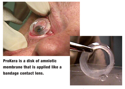
Biological bandage ring (ProKera, Bio-Tissue Inc.). This new device is comprised of a disk of amniotic membrane encircled in a polycarbonate band. It is applied like a bandage contact lens. Once the tissue dissolves, the surgeon removes the ring.
Tissue graft (AmnioGraft, Bio-Tissue Inc.). This is a square of amniotic membrane tissue affixed to filter paper. It is removed from the filter paper and placed over the defect. It adheres to the cornea on its own, or it can be secured with fibrin glue or sutures. If the defect goes deeper than the epithelium, multiple layers of tissue can be used to fill it.
Either preparation of the membrane dissolves in about one to two weeks, which is usually about as long as it takes for the defect to resolve, Dr. Woodard says.
Meanwhile, medical therapy should continue. In the case of a non-healing epithelial defect, for example, the patient should instill a topical antibiotic at least four times a day and artificial tears as frequently as possible to keep the cornea moist and healthy until the defect resolves.
After the procedure, the surgeon usually sees the patient the first day post-op and again in a week, Dr. Woodard says. Follow-up after this depends on how well the patient is healing. Usually, the patient returns to the optometrist after the membrane has dissolved and while the defect is in the process of resolving. So, when you see the patient again, assess visual acuity and monitor corneal integrity.
In rare cases, the membrane could slip off before the defect heals, Dr. Woodard says. Depending on how well the cornea is healing, the patient could either have another AMT or simply be monitored. Also, in rare cases, AMT may not effectively resolve the problem; these patients likely have stem cell disease and will need a stem cell transplant in addition to AMT.
