 |
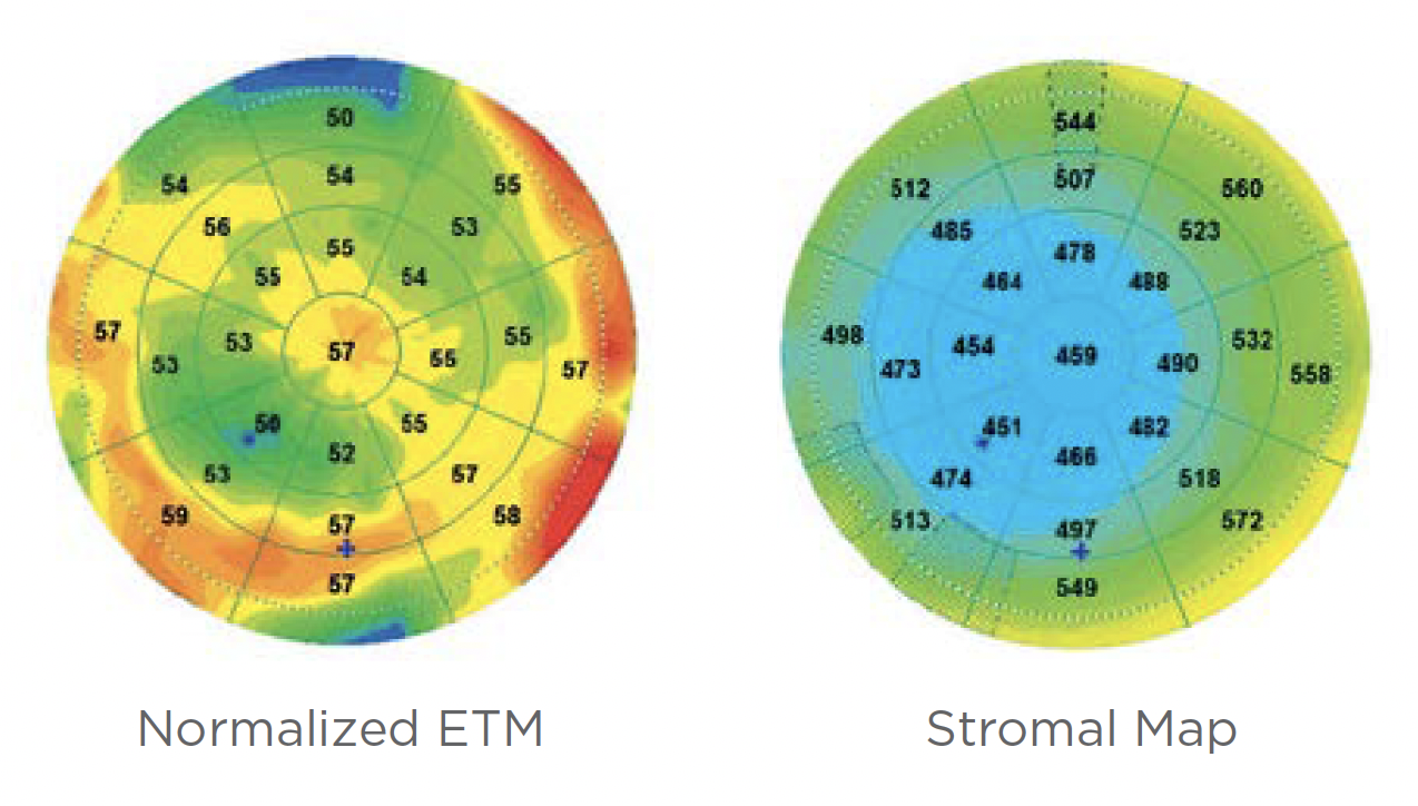
When compared with the Belin ABCD staging system, a newly developed OCT-based system that uses both epithelial (left) and stromal (right) thickness mapping showed staging agreement in 85% of normal cases and between 47% and 77% of keratoconus cases. Photo: Visionix. Click image to enlarge. |
As all clinicians know, catching keratoconus early enables earlier treatment with corneal crosslinking to help slow or halt disease progression. While detecting and grading the condition can sometimes be challenging, especially in cases of subclinical disease, a recent study developed and tested a new type of staging system that uses one of the most versatile tools optometrists can own—an OCT. The five-stage numerical system evaluates various ocular parameters on spectral-domain OCT (SD-OCT) and shows a strong agreement with existing systems. In addition, it is the first to take into account the epithelium and its sublayer stroma information, which led its creators to coin it the “STEP” staging system by combining the abbreviations for stroma (ST) and epithelium (EP).
To create the STEP system, the researchers conducted Scheimpflug tomography, air-puff tonometry and SD-OCT (RTVue-XR, Optovue) on 567 eyes of 567 patients including 331 with keratoconus of different stages and 236 normal controls. After analyzing the performance of each parameter in grading keratoconus, those that performed best were used to develop the new staging system.
The parameters with the highest sensitivity derived from the stroma and epithelium included stroma overall minimum thickness (sensitivity 90%, specificity 67%) and epithelium overall standard deviation (sensitivity 75%, specificity 78%). The STEP system strongly agreed with the two Belin ABCD staging systems; in normal patients, more than 85% of patients were in the same stage in all comparisons between the three systems. In keratoconus patients, the researchers reported that the staging agreement was still relatively high, ranging from 47% to 77%.
The team pointed out in their paper on the study, published in the Journal of Cataract and Refractive Surgery, that these outcomes “aligned well with previous studies using an alternative SD-OCT device (MS-39, CSO Italia, Firenze, Italy), where the authors demonstrated a good correlation between the degree of visual limitation (and KC severity staging) and stromal and epithelial thickness parameters.”
Though these data suggest the STEP system may be a reliable tool to stage keratoconus, the researchers point out a few limitations, one being that no progressive cases were included in the present study. Additionally, this analysis involved only one SD-OCT model, so further research will be needed to assess the system’s universal application.
“In conclusion,” the researchers wrote in their paper, “we propose a digital, automated, and comprehensive OCT-based keratoconus staging system that is compatible with existing keratoconus staging systems and offers additional clinical relevance. This system could be incorporated into daily clinical practice and in research, as it has the potential to help treatment decision-making and monitor keratoconus progression.”
Lu NJ, Hafezi F, Koppen C, et al. A novel keratoconus staging system based on optical coherence tomography. J Cat Refract Surg. [Epub ahead of print]. |
