 |
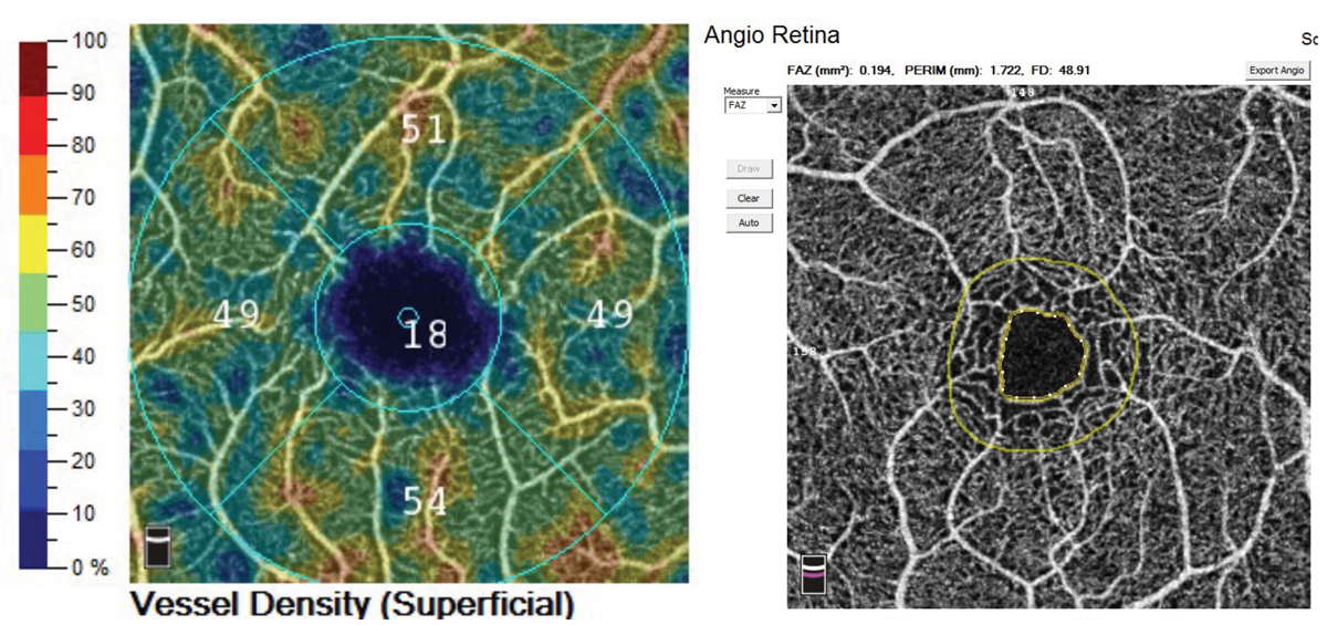
| The protocol used in this study demonstrated that the retinal capillary circulation around the FAZ was not uniform in healthy eyes. Photo: Jessica Haynes, OD. Click image to enlarge. |
The blood flow in the capillaries that form the foveal avascular zone (FAZ) is prone to stagnation in common retinal circulatory diseases such as DR and RVO, and the FAZ tends to enlarge over time. FAZ enlargement is considered an important clinical marker because of its significant associations with disease severity. A methodology called variable interscan time analysis was developed to detect abnormal blood flow in OCT-A devices by changing the interscan times to delineate relative blood flow velocity. OCT-A with longer interscan times revealed slower flow points in capillaries and more accurate visualization and morphology of microaneurysms in DR.
A recent study examined the retinal circulation status at the macula in healthy individuals using multiple interscan times. Extending the interscan times found new retinal capillaries with slow blood flow around FAZ. The researchers believed that such capillaries may be more likely to be more susceptible to retinal circulatory disturbance than the capillaries that did not require prolonged interscan times to be detected.
The study, conducted in Kyoto, involved OCT-A (Xephilio OCT-A1, Canon) scanning of a macular area measuring 4x4mm2 of 14 healthy eyes of 14 healthy volunteers (mean age 44) with no history or evidence of systemic and macular diseases. Interscan times were set at 7.6msec (the device’s default setting), 12.0msec and 20.6msec. Ten OCT-A images were acquired at each interscan time, and an averaged image was created. For each averaged OCT-A image obtained at the three times, the researchers defined the area surrounded by the innermost capillary ring as FAZ. They qualitatively evaluated the delineation of the capillaries consisting of the FAZ and quantitatively measured the FAZ area at each interscan time.
The extensions beyond the default both resulted in depiction of new capillaries around FAZ undetectable at the default interscan time. New capillaries were detected in 10 (71%) eyes at12.0msec and 11 (78%) eyes at 20.6msec. The researchers also found the FAZ areas to decrease significantly, as follows:
Interscan time | 7.6msec | 12.0msec | 20.6msec |
FAZ area | 0.334mm2 | 0.320mm2 | 0.319mm2 |
The research team noted that their protocol in this study may have the potential to enable a detailed assessment of the circulatory status in healthy eyes and eyes with retinal diseases. Further prospective studies with a greater number of participants and pathologic eyes will be required to confirm the findings of the present study.
“OCT-A has become the leading modality for the noninvasive evaluation of the retinal circulatory status,” the authors wrote in their paper.
Ishikura M, Muraoka Y, Nishigori N, et al. Macular retinal circulation in healthy eyes examined by optical coherence tomography angiography extended interscan time analysis. PLoS One. 2023;18(9):e0289896. |
