 |
|
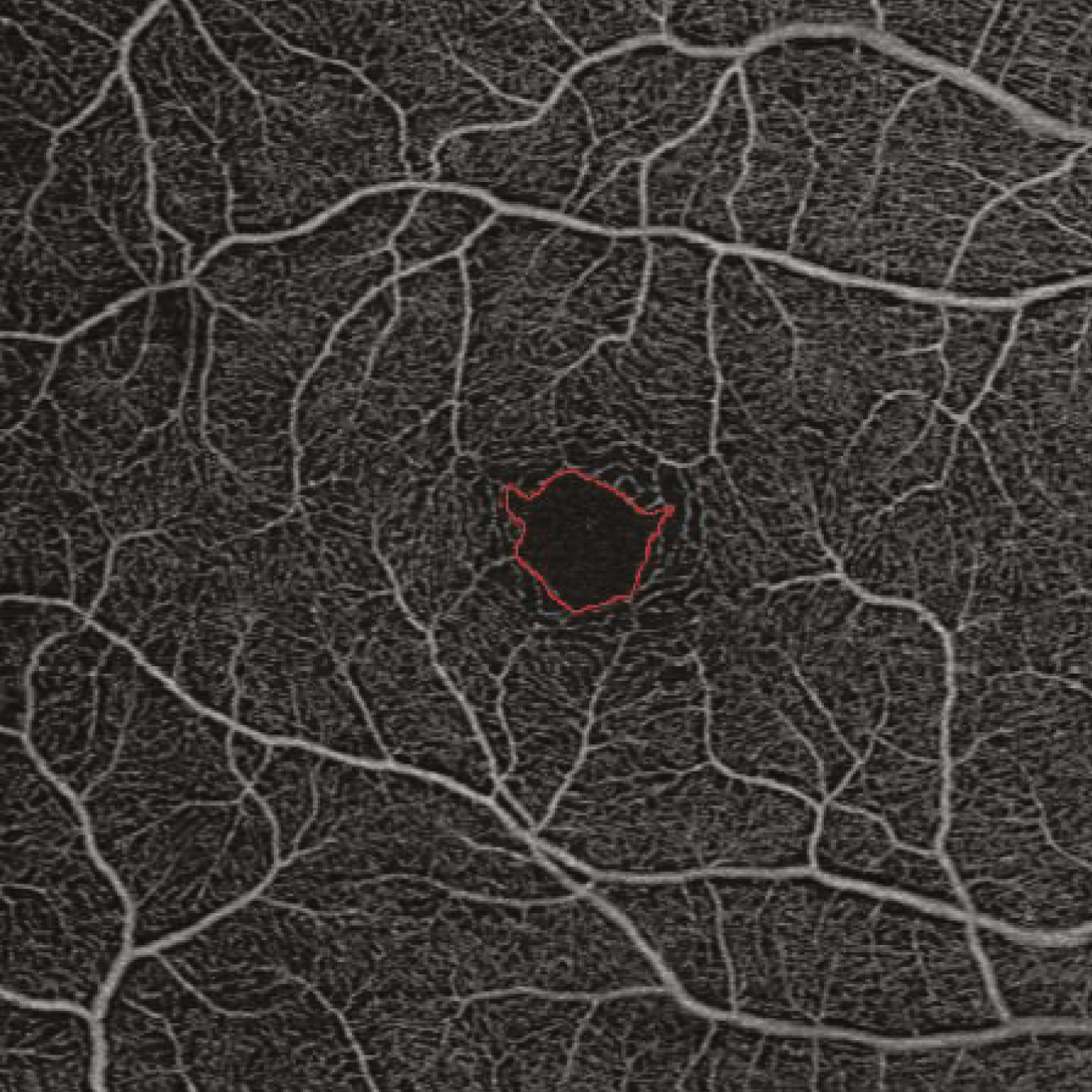
The added sensitivity provided by SS-OCT angiography may enable pre-clinical identification of diabetic eye disease, this study suggests. Photo: Vujosevic S, et al. Journal of Diabetes Research, 2019, 2547216.Click image to enlarge. |
New research suggests that macular retinal nerve fiber layer (RNFL) and ganglion cell-inner plexiform layer (GC-IPL) thickness are indicators for the early detection of diabetic retinal disease among type 2 diabetes mellitus (T2DM) patients who don’t have clinically observable retinopathy.
In this prospective, cross-sectional study, all eyes underwent swept-source optical coherence tomography (SS-OCT) angiography. The researchers performed a quantitative analysis of the acquired images that compared macular neural and microvascular alterations in T2DM patients without retinopathy (n=49) to age-matched controls (n=51). They measured the thickness of each individual retinal layer and evaluated macular vascular indices within different capillary plexuses.
Data showed that T2DM patients had a significant reduction in the mean macular thickness of the ganglion cell-inner plexiform layer (GC-IPL) and macular retinal nerve fiber layer (RNFL). Additionally, the study authors reported that macular full retinal thickness was significantly lower in diabetic eyes versus healthy controls. Subtle changes were observed in macular vascular skeleton density within the total capillary plexuses among T2DM patients.
“The study underscores the potential clinical utility of SS-OCT for the early detection of diabetic retinal disease in individuals with T2DM. The identification of retinal neurodegeneration through SS-OCT emerges as a potential precursor to subsequent microvascular changes,” the study authors noted in their recent American Journal of Ophthalmology paper. “These measurements exhibit promise as surrogate markers, enabling the stratification of patients with T2DM at an elevated risk of progressing to diabetic retinopathy.
“Looking ahead, SS-OCT emerges as a valuable diagnostic tool for the early identification of diabetic retinal disease, facilitating longitudinal assessments—especially crucial when neuroprotective interventions become available,” they stated. “Further investigations are warranted to elucidate the correlation and temporal evolution of structural and vascular changes in the development of diabetic retinal disease.”
Dai Y, Zheng D, Zhao J, et al. Macular Neural and Microvascular Alterations in Type 2 Diabetes Without Retinopathy: A SS-OCT Study. American Journal of Ophthalmology. February 28, 2024 [Epub ahead of print]. |
