 |
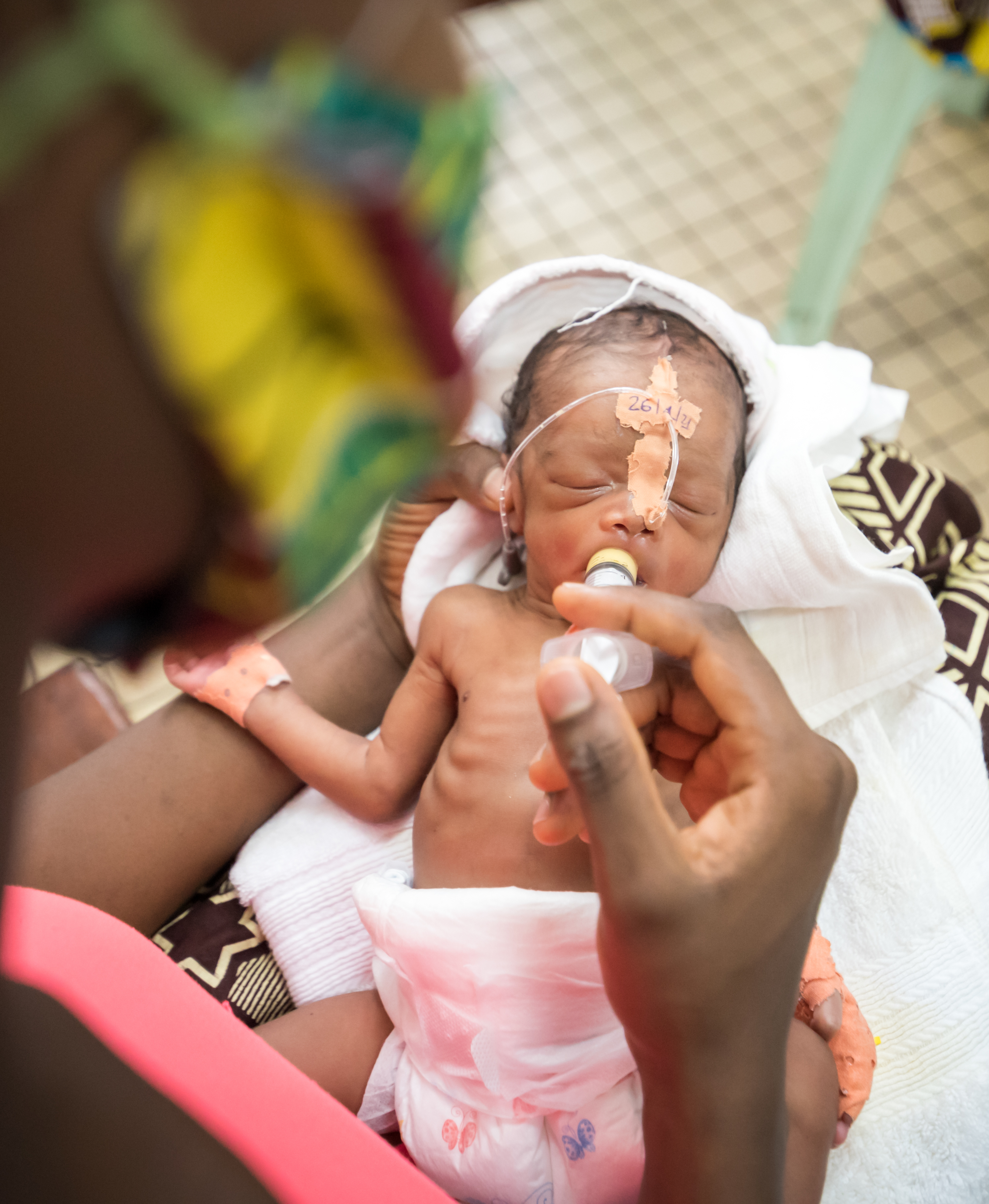
| While gestational age at birth and treatment for retinopathy of prematurity are risk factors for a thicker macula, researchers found no significant risk factors affecting the reduction of the ganglion cell-inner plexiform layer. Photo: Max Mbakop/Wikimedia Commons. Click image to enlarge. |
The ocular risks of premature birth are fairly well-known, as retinopathy of prematurity has been extensively studied. The ocular status in adulthood of these individuals, however, is less clear. For adults who had been premature at delivery, how have their retinas formed throughout adulthood whilst undergoing early life pathologies?
In a study to investigate central retinal morphology and retinal nerve fiber layer (RNFL) thickness in such individuals, researchers compared prematurely born young adults and term-born adults as controls. The premature group consisted of 59 individuals with a birthweight of less than or equal to 1,500 grams. Forty-four term born controls, all between the ages of 25 and 29, were assessed in the study. Researchers used OCT to evaluate retinal macular thickness, ganglion cell-inner plexiform layer thickness and retinal nerve fiber layer thickness.
Here are the results from the study: The central macular thickness in premature candidates was increased compared to the controls, with a mean change of 26.7µm. Even after excluding candidates with previous retinopathy of prematurity and those with neurological complications, the macular ganglion cell-inner plexiform layer was thinner for premature candidates (mean difference 3.84µm). This stayed true when researchers evaluated the candidates’ RNFL thickness. Even after excluding the aforementioned candidates with complications, the RNFL was thinner in premature candidates (mean difference 4.61µm) compared to the control. Researchers noted that both better visual acuity and contrast sensitivity were associated with thinner RNFL and thicker ganglion cell-inner plexiform layer.
Researchers concluded that macular and optic nerve morphology were influenced by premature birth compared to adult individuals following OCT assessments. Depending on the gestational age at birth and treatment for retinopathy of prematurity, this will have an effect on central macular thickness. Candidates who’ve been treated for retinopathy of prematurity had their peripapillary RNFL affected, and researchers noted that retinal sequelae remained through adulthood.
This study was unique in that it was conducted with formerly premature born individuals. “The prematurely born individuals have been followed since birth, and a majority of the control group has been followed from 10 years of age,” mentioned the researchers in their paper on the study. However, they did not perform OCT in childhood since it was not in clinical use at that time, which meant researchers could not include longitudinal comparisons.
Pétursdóttir D, Åkerblom H, Holmström G, Larsson E. Central macular morphology and optic nerve fibre layer thickness in youn adults born premature and screened for retinopathy of prematurity. Acta Ophthalmologica 2023;00:1-10. |
