A 63-year-old white female presented in the fall of 1998 for a routine primary eye care visit. She was taking Vaseretic (enalapril/hydrochlorothiazide, Merck), Synthroid (levothyroxine sodium, Abbott), and 20mg Nolvadex (tamoxifen, AstraZeneca) qd. She had a 10-year history of hypertension, and had abnormal thyroid studies for more than 20 years.

She also had breast cancer. About nine months before initial presentation, she underwent a complete right mastectomy followed by radiation therapy. She started on tamoxifen about four months prior to her initial presentation to me.
Her chief complaint at that time was decreased vision at distance and near, which had become more noticeable since her mastectomy. In her family history, she reported that her mother has glaucoma.
Diagnostic Data
Pupils were equal, round and reactive to light and accommodation with no afferent pupillary defect. Extraocular motilities were full. Best-corrected visual acuities were 20/20 O.U. through hyperopic astigmatic presbyopic correction.
A slit lamp examination of her anterior segments was unremarkable other than for fine SPK over the inferior one-third of her cornea, apparently dry eye related. She said that both eyes had been feeling drier over the past year. Applanation tensions were 20mm Hg O.D. and 21mm Hg O.S. Pachymetry was not obtained in the early visits.
Through dilated pupils, her crystalline lenses were clear O.U. Her cup-to-disc ratios were 0.65 x 0.65 in both eyes. The optic disc was slightly oval vertically, as was the cup. The neuroretinal rims were healthy. Maculae in both eyes were healthy with no foveal reflex. The vasculature was characterized by grade 1 atherosclerotic retinopathy.
Three weeks later, IOPs were essentially the same O.D. and O.S. Threshold visual fields were normal, but with a few paracentral points outside statistical threshold levels. SWAP (24-2) fields were normal O.D and O.S. Gonioscopy demonstrated 3+ open angles O.U., with normal trabecular pigmentation. Examination of the nerve fiber layer demonstrated a somewhat thinned NFL in the inferotemporal quadrant O.D. and O.S.
At that time, I elected to monitor the patient as a glaucoma suspect with ocular hypertension. She came in for follow-up exams approximately every six months.
In 2000, the patient presented with complaints of chemosis and increasing dry eye symptoms O.S. Within two weeks of the onset of these symptoms, the patient returned with diplopia, restrictive muscle function O.S. (left supraduction deficits), and 3mm of left-sided proptosis. MR imaging demonstrated classic findings associated with Graves orbitopathy, with significant muscle infiltration, O.S.> O.D. Thyroid function studies were normal except for elevated thyroid releasing hormone levels.
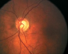
The patients optic disc was slightly oval vertically, as was the cup.
We obtained an endocrinology consult, and her Synthroid dosage was adjusted. The proptosis regressed over the next year and she retained some but not all EOM function. During the first six months of the orbitopathy, IOPs initially increased to 32mm Hg in both eyes, but settled to an average of 26mm Hg. Her optic nerves have remained stable. Threshold fields have also remained stable, although reliability of the left field was reduced primarily due to the inability of the left eye to fixate with relatively normal head posture. SWAP fields have demonstrated an increase in mean deviation.
Over the next two and a half years, I saw the patient at three-month intervals. IOPs have averaged 22mm Hg O.D. and 23mm Hg O.S. Pachymetry readings showed CCTs of 542m O.D. and 535m O.S. Nerves and NFL readings have remained stable. SWAP fields have shown a distinct increase in mean deviation, while standard threshold fields (white-on-white) have remained essentially stable.
Discussion
In patients developing glaucoma, SWAP field testing may demonstrate visual field abnormalities earlier than traditional white-on-white (WOW) threshold perimetry. Over the past five years though, this patient has had a distinct increase in mean deviations of SWAP fields, while the traditional WOW fields remained stable. That seems to indicate that this patient is converting to glaucoma, and medical intervention is necessary.
A recent study demonstrated that as the duration of tamoxifen use increases, so too do the effects on decreasing threshold levels in SWAP perimetry.1
Traditionally, tamoxifen is used for a five-year period, although some studies indicate benefits after just two years. It is used as both adjuvant therapy in breast cancer patients, as in the case of this patient, or in the management of advanced metastatic breast cancer. Tamoxifen is widely reported to induce corneal and lenticular changes, and also to induce retinopathy, with resultant changes in color vision and contrast sensitivity.
In this patients case, the change over time in the SWAP fields could very well be due to long-term tamoxifen use rather than actual conversion to glaucoma.
To muddy the waters even more, another study recently reported a higher incidence of OAG in patients with thyroid disease, in particular those taking thyroxine and those having undergone thyroid surgery.2 This study postulates that thyroid disease may be independently related to glaucoma.
One study indicates that the increase in SWAP mean deviations may be related to tamoxifen and not the development of glaucoma. Yet another study notes an increased correlation between glaucoma and patients on thyroxine. So how do you proceed?
In this case, until I have a clearer picture of conversion, I will continue to monitor this patient, and not medically intervene. But I will keep in mind the implications of recent studies, and clinically apply the specifics of those studies only after careful evaluation of the entire glaucomatous landscape.
1. Eisner A, Austin DF, Samples JR. Short wavelength automated perimetry and tamoxifen use. Br J Ophthalmol 2004 Jan;88(1):125-30.
2. Lee AJ, Rochtchina E, Wang JJ, Healey PR, Mitchell P. Open-angle glaucoma and systemic thyroid disease in an older population: The Blue Mountains Eye Study. Eye 2004 Jan 9 [Epub ahead of print]
