 |
|
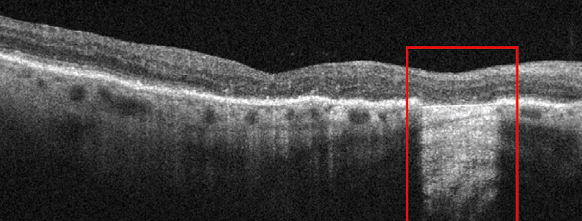
Greater attention to evidence of incomplete or complete (shown) RPE and outer retinal atrophy better identifies the status of GA patients. Photo: Anna Bedwell, OD. Click image to enlarge. |
A recent study, which explored whether the outer retinal layer thickness around geographic atrophy could help predict the annual enlargement rate of geographic atrophy, found a negative correlation between outer retinal layer thickness and annual geographic atrophy growth.
A total of 38 eyes from 27 subjects were included in this retrospective analysis of a prospective, observational case series. A swept-source OCT 6x6mm scan pattern was used to image eyes with geographic atrophy. The researchers measured geographic atrophy lesions with customized en face OCT images and calculated the annual enlargement rates. Choriocapillaris flow deficit analysis and retinal pigment epithelium (RPE) to Bruch’s membrane distance measurements were both conducted using the same set of eyes.
The outer retinal layer was defined and segmented from the inner boundary of the outer plexiform layer to the inner boundary of the RPE layer, according to the study authors who measured its thickness at different sub-regions around geographic atrophy.
The data showed a negative correlation between outer retinal layer thickness and geographic atrophy growth. While the study authors observed no correlation between the outer retinal layer thickness and the choriocapillaris flow deficit, they did report a significant negative correlation between the RPE to Bruch’s membrane distance and outer retinal layer thickness in the region immediately surrounding geographic atrophy.
“Due to the practical challenges of segmenting the outer retinal layer in a progressively degenerating disease, we propose that the RPE to Bruch’s membrane distance and choriocapillaris flow deficit measurements should suffice at this time as the baseline clinical biomarkers that predict about 60% of the annualized square-root enlargement rates of geographic atrophy as we explore for other anatomic biomarkers that may explain the remaining 40% of geographic atrophy growth,” the study authors noted in their recent paper.
Zhang Q, Shi Y, Shen M, et al. Does the outer retinal thickness around geographic atrophy represent another clinical biomarker for predicting growth? Am J Ophthalmol. August 20, 2022. [Epub ahead of print]. |
