 |
The location of structural progression was a key factor in whether an eye would experience pRNFL- or mGCIPL-progression first. Photo: Andrew Rouse, OD. Click image to enlarge. |
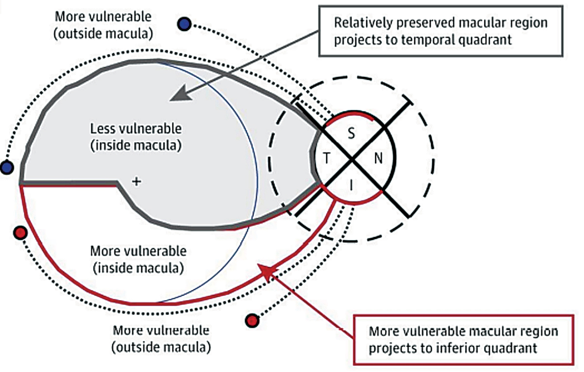
Structural data is a valuable clinical adjunct to visual fields when monitoring patients for glaucomatous progression. Researchers recently investigated whether optic disc changes measured by OCT influence the location at which functional changes are first detected—in the macular ganglion cell-inner plexus layer (mGCIPL) or peripapillary retinal nerve fiber layer (pRNFL), both of which are associated with visual field alterations. In their study, published in the Journal of Glaucoma, the group found that the cup-to-disc ratio could help predict where initial progression may occur.
The retrospective study included medical records of 282 eyes, 104 of which demonstrated structural progression in the mGCIPL (37 eyes) or pRNFL (49 eyes). The researchers observed significant differences between the two groups in terms of minimum mGCIPL thickness, pRNFL thickness, location of progression, and average and vertical cup-to-disc ratios.
Multivariate analysis showed that average and vertical cup-to-disc ratios were significantly associated with progression site. Additionally, they found that structural changes in the nferior area and superior vulnerability zone were significantly associated with RNFL-first progression.
“The cup-to-disc ratios and the location of the structural progression had a substantial influence on whether the initial structural progression was observed in pRNFL or mGCIPL,” the researchers wrote in their Journal of Glaucoma paper. “Our results showing that mGCIPL progression is more likely to be detected first in eyes with large vertical cup-to-disc ratios may help doctors detect progression earlier and, thereby, improve the prognosis in glaucoma patients.”
Park YM, Park JW, Bae H, et al. Optic nerve head morphology is associated with the initial location of structural progression in early open-angle glaucoma. J Glaucoma 2023. [Epub ahead of print]. |
