 Having taught at a major university for a combined total of 35 years, it’s not uncommon for us to get e-mails from colleagues with questions about unusual cases. One recent request from a former student went as follows:
“I have a patient here who is from Russia. Her brother will be visiting soon and she would like him to be seen by a retinal specialist. He has pathological myopia. The doctors in Moscow are recommending a ‘scleroplasty.’ They said he would ‘go blind’ in five years. Naturally, he is afraid and would like another opinion. Thank you for any information you can give me.”
Pathological myopia, also described in the literature as malignant, degenerative or progressive myopia, is a condition defined by refractive error in excess of -6.00D with an axial length greater than 26mm.1 In addition, the disease is characterized by “degenerative and progressive” changes involving a physical stretching of the sclera, choroid and retina.
The prevalence of progressive myopia varies greatly throughout the world. In the U.S., prevalence is roughly 2%; in other countries, it ranges from 1.7% to 3.3%.1,2 Certain populations can display greatly increased prevalence, however; the highest (24%) noted is in urban university students in Southeast Asia.3,4 Women tend to be affected more often than men, but this depends upon the population studied.2
History and Diagnostic Data

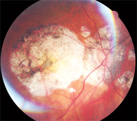
Key findings associated with progressive myopia can be seen on ophthalmic exam.
In virtually all cases, progressive myopia has a genetic predisposition, and elongation of the globe begins in early childhood.5 The precise etiopathogenesis of this disease is still unclear, despite much speculation. Papers published in the 1930s and 1940s implicated such things as squinting secondary to undercorrected refractive error and systemic calcium deficiency.6,7 More recent theories suggest that neurochemical processes trigger a signal cascade based on a visual feedback mechanism, which induces choroidal and scleral remodeling.8,9 Changes in the sclera’s extracellular matrix result in decreased durability, with excessive susceptibility to stretching. This may reflect systemic disorders of connective tissue metabolism.8,10
The key findings associated with progressive myopia can be seen on ophthalmoscopic examination of the posterior pole: myopic crescent; posterior staphyloma; flat, obliquely inserted discs; and patchy choroidal atrophy. Extensive vitreous syneresis and posterior vitreous detachment are typical. Peripheral retinal degenerations—lattice or snail-track, pavingstone and white without pressure—are also common. Additional findings occur with some variability and may include lacquer cracks, subretinal neovascular membrane, Fuchs’ spot (subretinal neovascularization with overlying RPE hyperplasia), retinal breaks and retinal detachments.11,12 Progressive myopia often accompanies systemic disorders such as Marfan’s syndrome, Ehlers-Danlos syndrome, albinism and premature birth.5
Management
The management of progressive myopia can be quite involved. When detected at an early age, most experts recommend full refractive correction (either via spectacles or contact lenses)—not undercorrection, which has been implicated in the development of greater amounts and rates of myopic progression.13,14
Unfortunately, no commercially available medications retard the process of axial elongation in progressive myopia, but at least one drug holds promise. The systemically-administered adenosine receptor antagonist 7-methylxanthine was shown to help normalize the growth of myopic eyes in a three-year study involving 107 children between the ages of eight and 13.15 Axial growth in the study group was reduced by as much as 22% vs. placebo.15 It is believed that 7-methylxanthine increases the content of collagen and proteoglycans in the sclera, as well as the diameter of collagen fibrils.16
Surgical intervention for progressive myopia is typically aimed at the specific pathological sequelae of the disease, such as subretinal neovascularization, peripheral retinal tears and retinal detachment.
Rarely discussed is a prophylactic procedure known as scleroplasty (sometimes referred to as “scleral strengthening” or “scleral reinforcement”), which is designed to help stabilize scleral extension and arrest myopic elongation of the globe. The procedure was pioneered some 80 years ago in pre-Soviet Russia, and during the subsequent 40 years, numerous alterations to the original technique were attempted.17,18 Ultimately, most experts in the Western world labeled this surgery as disappointing and without merit. Lacking conclusive evidence regarding the safety and efficacy of scleroplasty, this approach was widely abandoned in most regions of the world except in Eastern Europe and Asia.
But, a recent paper offers a possible rationale for resurrecting scleroplasty, in a technique called “posterior pole buckling.”19 This five-year study of 59 patients (ages 18 to 68) found that axial myopic progression was substantially diminished in eyes undergoing the buckling procedure when compared to fellow eyes, which served as a control group. In addition, there was a trend toward improvement in final visual acuity seen in the surgical group, while the acuity in control eyes diminished. Postoperative complications were low and non-sight-threatening, consisting primarily of temporary motility deficits, transient elevation of intraocular pressure and shallow choroidal effusions.
The authors concluded that posterior pole buckling possesses an acceptable safety profile and may be effective for the control of axial myopia, though additional prospective study is clearly necessary.
In a subsequent report published in the same journal, Russian physicians touted their experience with 183 patients undergoing scleroplasty for progressive myopia, with a follow-up period of up to 10 years.20 Although they did not share specific data for each patient, the group claimed that their procedure resulted in arrested progression of myopia in 95% of cases, as determined by refraction and axial length. The refraction decreased by 0.5D to 3.0D (mean: 1.3D) in 61.5% of cases, according to this report.
Meanwhile, a group in China reported similar success in a small study of 24 eyes with macular retinoschisis secondary to progressive myopia; in their series, 100% of subjects showed stabilization of refractive error, and 75% demonstrated improved visual acuity.21
Until preemptive medication or surgery becomes the norm in progressive myopia, we can only address the visual needs of these patients and continue to monitor them for complications. Those displaying evidence of subretinal neovascularization should be referred promptly for treatment with laser photocoagulation, photodynamic therapy or intravitreal injection of anti-VEGF therapies, such as bevacizumab.22
Patients with progressive myopia should be screened carefully and regularly (once or twice a year) for peripheral retinal degenerations, retinal breaks and detachments, and referred for treatment as indicated.
Advise these patients to avoid dangerous or jarring activities such as contact sports, bungee jumping or high-thrill amusement park rides. Such behavior can potentiate the risk for sight-threatening retinal damage
Thanks to Tracy Carpenter Sepich, O.D., M.S., of State College, Pa., for raising this intriguing issue.
1. Zejmo M, Formińska-Kapuścik M, Pieczara E, et al. Etiopathogenesis and management of high-degree myopia. Part I. Med Sci Monit. 2009 Sep;15(9):RA199-202.
2. Sperduto RD, Seigel D, Roberts J, Rowland M. Prevalence of myopia in the United States. Arch Ophthalmol. 1983 Mar;101(3):405-7.
3. Lin LLK, Shih YF, Hsiao K, et al. Epidemiologic study of the prevalence and severity of myopia among schoolchildren in Taiwan in 2000. J Formos Med Assoc. 2001 Oct;100(10):684-91.
4. Lin LLK, Jan JH, Shin YF, et al. Epidemiologic study on ocular refraction among children in Taiwan in 1995. Optom Vis Sci. 1999 May;76(5):275-81.
5. Zejmo M, Formińska-Kapuścik M, Pieczara E, et al. Etiopathogenesis and management of high myopia. Part II. Med Sci Monit. 2009 Nov;15(11):RA252-5.
6. Walker JP. Progressive myopia: a suggestion explaining its causation, and for its treatment. Br J Ophthalmol. 1932 Aug;16(8):485-8.
7. Burton EW. Progressive myopia: a possible etiologic factor. Trans Am Ophthalmol Soc. 1942;40:340-54.
8. Czepita D. [Contemporary views on the etiology, pathogenesis as well as treatment of school-age and progressive myopia] Klin Oczna. 1999;101(6):477-80.
9. McBrien NA, Gentle A. The role of visual information in the control of scleral matrix biology of myopia. Curr Eye Res. 2001 Nov;23(5):313-9.
10. Schaeffel F, Simon P, Feldkaemper M, et al. Molecular biology of myopia. Clin Exp Optom. 2003 Sep;86(5):295-307.
11. Saw SM, Gazzard G, Shih-Yen EC, Chua WH. Myopia and associated pathological complications. Ophthalmic Physiol Opt. 2005 Sep;25(5):381-91.
12. Pierro L, Camesasca FI, Mischi M, Brancato R. Peripheral retinal changes and axial myopia. Retina. 1992;12(1):12-7.
13. Chung K, Mohidin N, O’Leary DJ. Undercorrection of myopia enhances rather than inhibits myopia progression. Vision Res. 2002 Oct;42(22):2555-9.
14. Adler D, Millodot M. The possible effect of undercorrection on myopic progression in children. Clin Exp Optom. 2006 Sep;89(5):315-21.
15. Trier K, Munk Ribel-Madsen S, Cui D, Brøgger Christensen S. Systemic 7-methylxanthine in retarding axial eye growth and myopia progression: a 36-month pilot study. J Ocul Biol Dis Infor. 2008 Dec;1(2-4):85-93.
16. Cui D, Trier K, Zeng J, at al. Effects of 7-methylxanthine on the sclera in form deprivation myopia in guinea pigs. Acta Ophthalmol. 2009 Oct 23. [Epub ahead of print]
17. Shevelev MM. Operation against high myopia and scleralectasia with aid of the transplantation of fascia lata on thinned sclera. Russian Oftalmol J. 1930;11(1):107-10.
18. Snyder AA, Thompson FB. A simplified technique for surgical treatment of degenerative myopia. Am J Ophthalmol. 1972 Aug;74(2):273-7.
19. Ward B, Tarutta EP, Mayer MJ. The efficacy and safety of posterior pole buckles in the control of progressive high myopia. Eye (Lond). 2009 Dec;23(12):2169-74.
20. Balashova NV, Ghaffariyeh A, Honarpisheh N. Scleroplasty in progressive myopia. Eye (Lond). 2010 Jul;24(7):1303.
21. Zhu Z, Ji X, Zhang J, Ke G. Posterior scleral reinforcement in the treatment of macular retinoschisis in highly myopic patients. Clin Experiment Ophthalmol. 2009 Sep;37(7):660-3.
22. Montero JA, Ruiz-Moreno JM. Treatment of choroidal neovascularization in high myopia. Curr Drug Targets. 2010 May;11(5):630-44.

