 |
Q: A 21-year-old presented to my office stating that his right lid was “swollen” and he was seeing double at times. Where do I start with the workup?
A: “I would first investigate for an infectious/infiltrative process of the eye with the lid swelling,” says Leonard Messner, OD.
Orbital cellulitis, orbital pseudotumor and other infiltrative processes commonly produce eyelid edema with restriction of ocular motility and frequently affect the optic nerve to produce visual dysfunction, Dr. Messner says.
Of course, there may be more to the story. “If the involved lid is more of a ptotic vs. swollen lid, this changes the decision tree to explore the differentials related to ptosis associated with diplopia,” Dr. Messner says. “If the ptosis and motility deficits are ipsilateral, investigate for isolated or combined cranial neuropathy, such as oculomotor palsy or cavernous sinus syndrome, respectively.”
If the ptosis is associated with a contralateral motility deficit, this points strongly to myasthenia gravis (MG). “If the motility and eyelid dysfunctions do not obey the rules of neuroanatomy and vary over the course of the day, think MG,” says Dr. Messner.
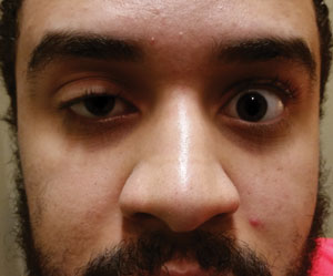 | |
| Acquired ptosis in one eye and a motility restriction in the other should be considered myasthenia gravis until proven otherwise. | |
 |
Diagnostic Tools
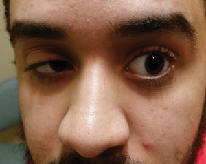
If you suspect neuromuscular disease, use your diagnostic toolbox to elucidate its exact etiology. For example, “Osher’s ‘peek sign’ is performed by asking the patient to gently close their eyelids as if they are sleeping,” says Dr. Messner. “Since concomitant orbicularis weakness is common with MG, it’s often easy to manually pry the lids apart.”
Ptosis and diplopia may not always occur together in MG. “Ptosis alone may be the first clinical presentation of ocular MG,” Dr. Messner says. “So, check all patients with ptosis for orbicularis weakness.”
You can also use Cogan’s lid twitch. “I ask the patient to look in a slightly downward position for 10 to 20 seconds, then redirect their gaze to primary position. With the redirection, the upper lid will typically overshoot or twitch before coming down to its normal position,” Dr. Messner explains.
An ice pack is also an effective MG diagnostic tool. Dr. Messner uses crushed ice in a surgical glove applied to the eye with closed lids for several minutes. Improvement of the ptosis is highly suggestive of MG. He also takes facial photos before and after to check for any signs of improvement.
Historically, 50% to 80% of ocular MG cases convert to generalized MG, with later-in-life diagnosis as a risk factor for conversion, Dr. Messner says, with recent research showing a conversion rate of 21%.1 He is careful to note that ocular myasthenia gravis is not distinct from generalized myasthenia gravis. “It is incorrect to think of ocular myasthenia and generalized myasthenia as distinct disease processes; rather, ocular MG represents an early form of the disease,” notes Dr. Messner.
It is important to question patients as to the presence of other bulbar signs (difficulty chewing, swallowing and breathing), as these are also common with MG, yet are often overlooked or misdiagnosed, Dr. Messner says.
“Many patients complain of difficulty swallowing, with the build-up of phlegm in their throat owing to paralysis of their laryngeal muscles,” Dr. Messner says. He has also seen patients with breathing problems initially attributed to sinus disease. “If I suspect an individual of ocular MG, I order acetylcholine antibody titers along with a chest CT to rule out thymoma, which is present in about 15% of the MG cohort.”
Treatment
The initial goal is to effectively treat the underlying disease process. Since anticholinesterase agents do not treat its autoimmune etiology, early treatment with prednisone is the evolving trend, according to Dr. Messner. Immunosuppresant therapy can reduce the rate of conversion to 10.5%.1
“While pharmacotherapy is much more effective than in the past, many individuals will continue to have problems with persistent ptosis and diplopia,” says Dr. Messner. Optometric care may include ptosis crutches or eyelid taping. For diplopia during the acute and subacute stages, Dr. Messner suggests unilateral occlusion and prism therapy once stable.
1. Nagia L, Lemos J, Abusamra K, et al. Prognosis of ocular myasthenia gravis. Ophthalmol. 2015 April 16. [Epub].
