 A 67-year-old white female presented to the office as a new patient in the summer of 2007 after recently moving to the area. Her medical history was significant for hypertension, hyperlipidemia and sleep irregularities. Her medications included Diovan (valsartan, Novartis), atenolol, Questran (cholestyramine, Bristol-Myers Squibb) and Xanax (alprazolam, Pfizer) h.s. p.r.n. She also took over-the-counter ibuprofen as needed for aches and pains. She reported no allergies to medications.
During this initial visit, the patient mentioned that her previous eye care provider had been following her every six months because of “high pressures.”
Diagnostic Data
Best-corrected visual acuity was 20/20 O.D. and O.S. through mildly hyperopic astigmatic and presbyopic correction. Pupils were equal, round and reactive to light and accommodation with no afferent defect. Extraocular motilities were full in all positions of gaze.
Applanation tensions measured 23mm Hg O.D. and 24mm Hg O.S. at 3:15 p.m. Pachymetry readings were 579μm O.D. and 585μm O.S.
Slit lamp examination of her anterior segments was unremarkable, except for mild guttata in both eyes. Anterior chamber angles were open and estimated by Van Herick’s to be grade IV O.U.
Upon dilation, her crystalline lenses were characterized by incipient nuclear lens changes consistent with her age. Multiple vitreous floaters were present bilaterally.
I estimated her cup-to-disc ratios to be 0.55 x 0.60 O.D. and 0.60 x 0.60 O.S. Both maculae were characterized by loss of foveal reflexes and mild retinal pigment epithelium granulation, also consistent with her age. Her retinal vasculature was remarkable for bilateral and symmetric mild arteriosclerotic retinopathy. Her peripheral retinas were remarkable for scattered areas of pavingstone degeneration with no holes, tears or tractional phenomena O.U. I asked her to return in approximately six weeks for field studies, gonioscopy and HRT imaging so that I could gather more baseline information.
In the interim, I requested copies of her records from her previous eye doctor; in reviewing them, I noticed that her IOP ranged from 20mm Hg to 27mm Hg O.D. and O.S. at various visits. Further, threshold visual fields were all normal and optic nerve cup-to-disc ratios were judged slightly smaller than my estimates. Also, she had been compliant with her six-month follow-up schedule.
When the patient returned six weeks later for a morning appointment, her visual fields were reliable with no early glaucomatous defects. At 9:00 a.m., her IOP was 25mm Hg O.D. and O.S. Gonioscopy demonstrated open angles 360° O.U., with a flat, normal iris approach. Imaging of the optic nerves confirmed my earlier cup-to-disc measurements, with normal Moorfields indices in all sectors of the optic nerve.
I informed the patient of the findings and asked her to return every six months for evaluation of her optic nerve stability.
In late 2009, the patient presented for a scheduled follow-up visit, at which time her fields and nerve evaluations were unchanged from previous visits, but IOP readings were 29mm Hg O.D. and 31mm Hg O.S. On further questioning, the patient had recently discontinued a short course of oral steroids for acute bronchitis. I explained that her somewhat elevated IOP readings were possibly due to the recent steroid use, and asked her to return for a brief follow-up in one month, at which time her IOP had returned to the mid-20s.
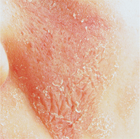
This patient’s moderate but body-wide dermatitis required oral steroids, which elevated her intraocular pressure.
In spring 2011, the patient presented on an urgent basis to have her IOP checked. She was concerned because she had been having a moderately body-wide dermatitis. After several treatment regimens, her dermatologist mentioned putting her on oral steroids to quiet the dermatitis, and she was concerned about the impact that could have on her IOP.
Apparently sensing anxiety on the part of the patient, the dermatologist deferred the oral steroids pending my approval. At this visit, which was before the oral steroids were initiated, her IOP was 24mm Hg O.D. and 26mm Hg O.S.
I informed the patient that her recalcitrant dermatitis would likely respond to oral steroids, and that she should follow through with the dermatologist’s recommendation. Though I believe the patient understood, I could tell she was anxious about taking the steroids, so I asked her to return in 30 days to have her pressures evaluated. She was very relieved by this request.
The patient returned as scheduled in 30 days. She was on a tapering dose of 10mg of prednisone b.i.d. Her IOP readings were 32mm Hg O.D. and 33mm Hg O.S. All other findings were unchanged from earlier visits.
Would you treat these pressures?
Discussion
Clearly, the patient was a steroid responder. In all likelihood, her IOP may have actually been higher a week or two prior to me seeing her. But the question remains, do these elevated pressures need to be reduced?
Several factors need to be considered in answering this question:
- Is the patient actually a steroid responder, or is she an ocular hypertensive patient whose IOP simply has risen?
- Is the steroid responder taking topical or oral steroids?
- How long will the patient be treated with steroids? And how much of the medication is she taking?
- How much of a steroid response would she typically have? Mild? Significant?
Of course, the answers to these questions will vary from patient to patient, ultimately depending on what condition is being treated. For example, some dermatological conditions may require a short-term (one- to three-week) steroid course, while other rheumatologic or respiratory conditions may require continuous steroid usage. Sometimes, we’ll put a patient on a topical steroid to manage some ocular inflammation, and that course of topical steroids may last days or months.
But the biggest factor that must be considered in managing a steroid responder is the severity of their optic nerve disease, and ultimately, the frailty of her optic nerves. In general, patients with healthier optic nerves can withstand elevations in intraocular pressure better than those with more fragile optic nerves. Keep in mind that many glaucoma patients will be steroid responders, and as such, these patients may already have optic nerve damage to one degree or another. Is the glaucoma patient rather young with minimal optic nerve changes, or is the state of the optic nerve such that there is more structural or functional damage already present, and therefore more susceptible to further damage?
In reality, all of these issues come in to play. For example, a patient with severe neuroretinal rim damage who is a steroid responder may in fact need IOP-lowering agents even if he is only going to be on the steroids for a short time. Likewise, a patient with little to no nerve damage may need to be on IOP-lowering agents especially if he is a profound responder and will be on steroids for an extended period of time.
Your decision whether to initiate IOP-modifying agents essentially boils down to your assessment of the ability of the optic nerve to NOT undergo deterioration while the IOP is elevated. This may involve assessing how high IOP actually goes before initiating therapy, or it may involve determining that the risk to the optic nerve is such that therapy is necessary prior to a rise in IOP. There is no set management plan for these patients because so many factors need to be considered, and these considerations vary from patient to patient.
In this patient’s case, although I thought that her optic nerves could handle a short duration of elevated IOP without damage, I also thought it was best to temporarily initiate IOP-modifying agents primarily due to both the level of IOP elevation, AND her personal heightened level of anxiety upon hearing her pressures were now in the low 30s. I asked her to begin 0.5% timolol 1 drop O.U. q.a.m. It was both an IOP management decision as well as a patient management decision to initiate this therapy.
As her oral steroid usage was discontinued over the next several weeks, her IOP declined as well. Before discontinuing the timolol, her IOP measured 19mm Hg O.D. and 20mm Hg O.S. After she discontinued it, her IOP was back in the mid-20s range, which is consistent for her.
This column is dedicated to the memory of Dr. Kevin Bell, of Wake Forest, N.C. The epitome of a great optometrist, Dr. Bell passed away in September.
