
Hypertension (HTN), or chronic abnormally high blood pressure, is one of the most prevalent diseases worldwide. It affects nearly 50 million Americans and two-thirds of the population above age 65.1,2 These numbers will likely increase as the American population ages.
But, HTN isnt always obvious, and sometimes a change in ophthalmic vasculature may be the key sign in the diagnoses of HTN. So, optometrists play a key role in comanaging hypertensive patients.
Diagnosis and Categorization
The point at which chronic elevated blood pressure becomes a diagnosis of hypertension is not always clear cut. For years, hypertension was defined as blood pressure greater than 140/90mm Hg.
Although the exact definition of hypertension is still ambiguous, the Joint National Committee (JNC) of Prevention, Detection, Evaluation, and Treatment of High Blood Pressure has established guidelines for diagnosis (see Guidelines for Diagnosing Hypertension").1 JNC Report 7 defines optimal blood pressure as a measurement of less than 120/80mm Hg.1 But, multiple variables must be considered, such as a noticeably elevated blood pressure reading on at least two chronologically separate visits.
Guidelines for Diagnosing Hypertension1 The following guidelines were published by the Joint National Committee of Prevention, Detection, Evaluation, and Treatment of High Blood Pressure: Pre-hypertensive: 120mm Hg to 139mm Hg systolic; 80mm Hg to 99mm Hg diastolic. Stage 1 hypertension: 140mm Hg to 159mm Hg systolic; or 90mm Hg to 99mm Hg diastolic. Stage 2 hypertension: 160mm Hg or greater systolic; or 100mm Hg or greater diastolic.
Hypertension is categorized as essential or secondary. Most cases are essential, diagnosed in the absence of any causative factor.3 Only 5% of cases are classified as secondary to an identifiable cause.3
Normal blood pressure: less than 120mm Hg systolic; less than 80mm Hg diastolic.
The pathogenesis of HTN is complex. Genetic predisposition, excess salt intake and adrenergic tone all play key roles in the development of HTN.3 But, other factors, such as obesity and race, have also been shown to influence the development of the disease. Blacks have an increased risk of developing HTN; the prevalence of hypertension among blacks is 33.5% vs. 28.7% in whites.4 Researchers have speculated that socioeconomic factors and lifestyle may contribute to this increased prevalence of HTN among blacks.5 Hypertension is insidious among Americans and is a leading cause of annual morbidity and mortality associated with cerebrovascular or cardiovascular disease.6 Persistent high blood pressure leads to organ damage and affects the brain, heart, kidneys and eyes.3
Ocular Manifestations of Hypertension Anterior ischemic optic neuropathy Central or branch retinal artery Central or branch retinal vein occlusions (CRVO or BRVO) Choroidal infarction Cranial nerve palsies Progression of diabetic retinopathy Glaucoma Hypertensive retinopathy Idiopathic polypoidal choroidal vasculopathy (IPCV) Macroaneurysms Ocular ischemic syndrome Subconjunctival hemorrhage Transient visual obscurations
Myriad ophthalmic vascular changes occur in response to both acute and chronic elevated blood pressure, and often affect the retina and choroid (see Ocular Manifestations of Hypertension).7 Because ophthalmic vascular changes may be warning signs of organ damage associated with HTN, optometrists play a critical role in diagnosing and comanaging hypertensive patients.
occlusion (CRAO or BRAO)
Hypertensive retinopathy (HTR) is the most common ocular manifestation of HTN. Arterial occlusion and ocular ischemic syndrome, which are often associated with cardiac and carotid disease, can also result from hypertension. Other common secondary ocular manifestations include choroidal infarctions, vein occlusion and retinal arterial macroaneurysm (RAM).7
Hypertensive Retinopathy
The term hypertensive retinopathy was first introduced in 1930.8 Since then, studies have reported that hypertensive patients have a 50% to 80% chance of developing HTR.9 Also, patients with signs of HTR are significantly more likely to have high blood pressure as well.9 Clinically, the initial signs of HTR are observed as alterations in the retinal microvasculature.
Several classification systems have been used to describe the continuum of microvasculature changes that occur in patients with HTR. One such system defines it as mild, moderate and malignant stages of hypertensive retinopathy.10
But, the most widely accepted classification is the Keith-Wagener-Baker system, which categorizes four stages of HTR (see Keith-Wagener-Barker Hypertensive Retinopathy Classifications).9,11
Keith-Wagener-Barker Hypertensive Retinopathy Classifications11 Stage 1: Mild retinal vascular changes (generalized arteriolar narrowing). Stage 2: Moderate to severe retinal vascular changes (arteriovenous crossing changes). Stage 3: Stage 1 and 2 findings, plus cotton-wool spots, retinal hemorrhages and exudates.
Stage 1. Mild generalized arteriole sclerosis with increased arteriole light reflex, secondary to thickening within the blood vessel wall. This presentation typically signifies a chronic hypertensive change.13 These changes canand dooccur in the normal elderly population.14
Stage 4: Stage 3 findings, plus associated optic nerve head swelling and macular star formation.
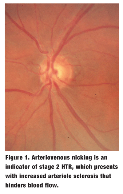
Stage 2. Moderate to severe retinal vascular changes are synonymous with decrease in lumen size and hyalinized arterial walls, which may lead to arteriovenous nicking, a pathognomonic feature of HTR (figure 1). At the arteriovenous crossing, the artery typically lies over the vein, sharing the same outer sheath. With increased arteriole sclerosis, the vein becomes susceptible to compression, which hinders normal blood flow.
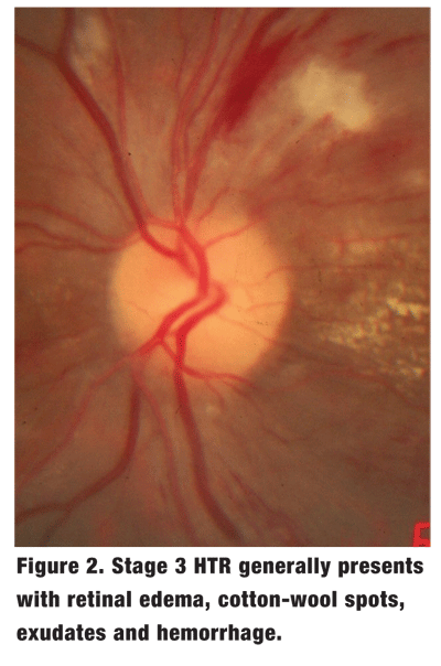
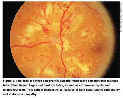
Stage 3. In this stage, acute elevation in blood pressure causes a breakdown of the blood-retina barrier. Contributing signs include retinal edema, cotton-wool spots, exudates and hemorrhage (figure 2). These microvascular changes are often difficult to distinguish from diabetic retinopathy (figure 3).
Unlike diabetic retinopathy, HTR usually exhibits a drier retina associated with more cotton-wool spots and less exudates and/or hemorrhages. Also, the predominant retinal hemorrhages associated with HTR are flame-shaped hemorrhages rather than dot-and-blot hemorrhages.
Stage 4. The most advanced stage of HTR is known as malignant hypertension. Malignant hypertension is accelerated high blood pressure that consists of a systolic pressure higher than 200mm Hg and a diastolic pressure higher than 140mm Hg. This serious increase in blood pressure is often associated with morbidity and/or mortality secondary to stroke, myocardial infarction, and renal and heart failure. Patients often present with complaints of decreased vision, headaches, diplopia, scotomas and/or photopsias. This stage is characterized by the signs of stage 3 HTR plus optic nerve head swelling and macular edema (macular star).
Currently, treatment for patients with stage 1, 2 or 3 HTR includes close observation, management of high blood pressure and regular dilated fundus exams. Malignant HTN, however, is a clinical emergency that warrants immediate referral for proper anti-hypertensive treatment. The reported three-year survival rate in patients with malignant hypertension is 6%, vs. 70% for those with stage 1 HTR.11 Fortunately, fewer than 1% of HTR cases are associated with malignant hypertension, primarily because it does not typically occur in treated hypertensive patients.9 Although the incidence of malignant HTN is rare, it can present in association with secondary causes of hypertension.
Of note, a study by Sohan Singh Hayreh, M.D., suggests that higher blood pressure is not always a prognostic marker for the advancement to the more severe stages of HTR.12
 Hypertensive Choroidopathy
Hypertensive choroidopathy manifests less often than HTR. Unlike the retinal vasculature system, the choriocapillaris are not autoregulated. They are thin with a high degree of fenestration, which makes them susceptible to severe acute rise in blood pressure.
Hypertensive choroidopathy is a sign of choroidal vascular insufficiency, resulting in choroidal and overlying retinal pigment epithelium (RPE) infarction. Focal ischemia within the choroidal vasculature results in occlusion of the choriocapillaris and leads to damage of the overlying RPE.
Two common manifestations of hypertensive choroidopathy are Elschnigs spot and Siegrists streak. Elschnigs spots are typically yellow circular lesions with associated hyperpigmentation, while Siegrists streaks are linear pigmented areas that run along sclerotic choroidal vessels.
Also, serous retinal detachment may occur in conjunction with hypertensive choroidopathy. Disruption of the RPE leads to a breakdown of the retinal blood barrier, which results in subretinal fluid accumulation.
 Hypertensive choroidopathy predominantly affects younger patients with severe acute hypertension.15 If you see any signs of this, refer the patient as soon as possible for further treatment.
Unlike diabetic retinopathy, there is no set or generally accepted system of classification for common posterior segment manifestation of hypertensive. So, it is often best to describe direct ocular manifestations based on the funduscopic changes, using angiopathy, retinopathy, choroidopathy and optic neuropathy as the main categories.
Vein Occlusions
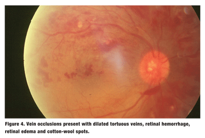
Retinal vein occlusions are typically associated with vascular disease. HTN is one of the most common of these. It contributes to thrombosis, which in turn leads to vein occlusion. Some 50% of branch retinal vein occlusions (BRVOs) are linked to HTN.16,17
The clinical picture of retinal vein occlusion is a middle-aged patient with complaints of acute vision loss. The retinal characteristics of vein occlusions include dilated tortuous veins, retinal hemorrhage, retinal edema and cotton-wool spots in response to back flow of blood (figure 4). Optic nerve head swelling and macular edema may also be present.
When the occlusion site is at the level of the central retinal veincentral retinal vein occlusion (CRVO)the presentation affects all four quadrants of the fundus. But, when the occlusion site is at the level of one of the major branchesBRVOonly the retina and vasculature at the distribution of the involved vein is affected.
The clinical picture may further vary if the vein occlusion is ischemic or non-ischemic. Ischemic vein occlusions have a greater likelihood of retinal non-perfusion, which increases the chances of neovascularization. Complications of neovascularization include neovascular glaucoma, tractional retinal detachment and vitreous hemorrhage. When managing vein occlusions, remember it is critical to treat the underlying disease. In addition, close follow-up for progression and development of complications is a necessity.
Treatment options for neovascularization and macular edema vary depending on the location and severity of each case. Current options include steroid injection, laser photocoagulation, anti-vascular endothelial growth factor agents, and surgical therapy.18
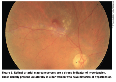
Fifty percent to 75% of presenting retinal arterial macroaneurysms (RAM) are associated with HTN. Specifically, HTN contributes to vessel wall weakening and leads to aneurysm formation.19,20
A RAM is an acquired localized round dilation that balloons over a major retinal arterial branch (figure 5). Typically, it has a unilateral presentation and affects women between the ages of 50 and 80 who have a systemic history of hypertension. Associated leakage in the form of exudation, retinal edema and hemorrhage may also be observed.
There are two distinct forms of RAMs. A fusiform RAM is spindle shaped, while a saccular RAM is shaped like a small sac.21 The saccular form is more likely to result in hemorrhaging, while the fusiform type is typically associated with exudation.
Also, the presentation of either form of RAM is unpredictable. It may present as a benign aneurysm, or it may be associated with variable layers of retinal hemorrhage, including vitreous hemorrhage.
Furthermore, weakening of the macroaneurysm wall may result in associated retinal edema and exudation in a circinate pattern. Although many patients initially are asymptomatic, macular involvement often leads to visual impairment.
Due to its variable presentation and complications, suspect a RAM whenever you observe localized isolated leakage over a retinal artery bifurcation. Angiography aids in the diagnosis, showing hyperfluorescence filling of the RAM, with possible associated leakage and microvascular anomalies.
Because the bleeding typically results in spontaneous aneurysmal thrombosis and vessel sclerosis, asymptomatic or non-leaking RAMs should be monitored closelyevery six months.22
Non-leaking RAMs near the macula, or leaking RAMs that do not threaten the macula require even closer monitoring.
Treatment is considered for a leaking RAM near the macula and persistent or progressive RAMs, particularly if a pulsating arterial wall is noted. A pulsating RAM indicates a thin wall, which is more likely to rupture.
Treatment options include photocoagulation laser treatment and Nd:YAG photodisruption.23-25 Photocoagulation laser treatment seals the leaking aneurysm. On the other hand, Nd:YAG photodisruption assists absorption of the associated hemorrhage by creating a slit on the retinal surface that allows the hemorrhage to drain into the vitreous cavity. This process speeds up the recovery of vision and prevents irreversible damage associated with complications and longstanding toxicity of hemorrhage to the retina. Currently, all RAM treatments are controversial with variable results.23-25
Management of HTN
At this time, about 30% of American hypertensive patients are unaware of their condition.4 Optometrists must be vigilant in looking for the acute and chronic ocular signs of hypertension, which may help to correctly diagnose these otherwise-asymptomatic patients.
Besides an ocular health evaluation, regular in-office blood-pressure monitoring is imperativein every comprehensive eye exam. To obtain accurate measurements, make sure to use the correct cuff size and correct cuff placement, and do not allow patients to smoke or consume caffeine before taking a reading.26
Ocular manifestations of hypertensive retinopathy may be reversible with control of the high blood pressure. Close monitoring every three to six months should be initiated at the early or moderate stages of HTR, in order to evaluate for resolution or progression.
The primary goal when managing the ophthalmic manifestations associated with hypertension is to establish and control the underline etiology. So, HTR is usually comanaged with the patients primary-care practitioner to normalize the blood pressure.27
Patients who have HTR warrant a thorough medical workup and may need to begin anti-hypertensive therapy.28,29 Hypertensive neuropathy is also reversible once the malignant hypertension is under control, and early treatment generally means better prognosis.
A patient who presents with malignant hypertension is at grave risk for further systemic complications and needs to be referred for urgent medical intervention. Be sure to convey to your patient the serious nature of the condition. Note: Because the patients end organ tissues have adapted to a malignant pressure, it is wise to decrease pressure slowly over a period of time to allow adjusted autoregulation to a lower perfusion.27 Other secondary ocular manifestations associated with HTN, such as vein occlusions and macroaneurysms, should be treated and monitored accordingly.
Hypertension is one of the most common medical conditions in the
Optometrists must become familiar with the variety of ophthalmic conditions associated with HTN, because early recognition of such changes is imperative for appropriate diagnosis and prompt intervention. There are numerous patients who are undiagnosed or under-treated and are thus at risk for all the consequences associated with HTN.
Whenever you encounter a patient who has ocular manifestations of HTN, but an otherwise unremarkable medical history, have the patient evaluated for undiagnosed HTN. Additionally, whenever a patient with known HTN manifests hypertensive ocular signs, you must effectively communicate with the patients primary-care physician, cardiologist or internist, since this may be evidence that the patients condition is not well controlled.
The optometrist plays a critical role in the diagnosis and management of patients with HTN. Early diagnosis of ocular manifestations associated with HTN may lead to more appropriate management and timely referral. By performing a comprehensive eye examination, which includes blood pressure evaluation, we can affirm the role of the optometrist as a primary heath-care provider.
Diana L. Shechtman, O.D., is an associate professor of optometry at
1. The 7th report of the Joint National Committee on Prevention, Detection, Evaluation, and Treatment of High Blood Pressure. December 2003. Available at: www.nhlbi.nih.gov/guide lines/hypertension/jncintro.htm (Accessed October 24, 2006).
2. Franklin SS, Jacobs MJ,
3. Sharma S, Kortas C. Hypertension. July 2006. Available at: www.emedicine.com/med/ topic1106.htm (Accessed October 12, 2006).
4. Hajjar I, Kotchen TA. Trends in prevalence, awareness, treatment, and control of hypertension in the United States, 1988-2000. JAMA 2003 Jul 9;290(2):199-206.
5. Wong TY, Klein R, Duncan BB, et al. Racial differences in the prevalence of hypertensive retinopathy. Hypertension 2003 May;41(5):1086-91. Epub 2003 Mar 24.
6. Graves EJ, Kozak LJ. National hospital discharge survey: Annual Summary, 1996. Vital Health Stat 13. 1999 Jan;(140):i-iv,1-46.
7. Schubert HD. Ocular manifestations of systemic hypertension. Curr Opin Ophthalmol 1998 Dec;9(6):69-72.
8. Fishberg AM, Oppenheimer BS. The differentiation and significance of certain ophthalmoscopic pictures in hypertensive disease. Arch Intern Med 1930;46:901-20.
9. Klein R, Klein BE, Moss SE, et al. Hypertension and retinopathy arteriolar narrowing, and arteriovenous nicking in a population. Arch Ophthalmol 1994 Jan;112(1):92-8.
10. Wong TY, Mitchell P. Hypertensive retinopathy. New Engl J Med 2004 Nov 25;351(22):2310-7.
11. Keith NM, Wagener HP, Barker NW. Some different types of essential hypertension: their course and prognosis. Am J Med Sci 1974 Dec;268(6):336-45.
12. Hayreh S, Servais G, Virdi P, et al. Fundus lesions in malignant hypertension: III. Arterial blood pressure, biomechanical, and fundus changes. Ophthalmology 1986 Jan;93(1):45-59.
13. Sharrett AR, Hubbard LD, Cooper LS, et al. Retinal arteriolar diameters and elevated blood pressure: The Atherosclerosis Risk in Communities Study. Am J Epidemiol 1999 Aug 1;150(3):263-70.
14. Leishman R. The eye in general vascular disease: hypertension and arteriosclerosis. Br J Ophthalmol 1957 Nov;41(11):641-701.
15. Bourke K, Patel MR, Prisant LM. Marcus DM. Hypertensive choroidopathy. J Clin Hypertens 2004 Aug;6(8):471-2.
16. Rath EZ, Frank RN, Shin DH, Risk factors for retinal vein occlusions. A case-control study. Ophthalmology 1992 Apr;99(4):509-14.
17. The Eye Disease Case-Control Study Group. Risk factors for branch retinal vein occlusion. Am J Ophthalmol 1993 Sep 15;116(3):286-96.
18. Shechtman D. Intervene against the interruption of blood flow. Second Annual Guide to Retinal Disease. Rev Optom 2005 Nov;142(9)Suppl:13-17.
19. Abdel-Khalek MN, Richardson J. Retinal macroaneurysm: Natural history and guidelines for treatment. Br J Ophthalmol 1986 Jan;70(1):2-11.
20. Panton RW, Goldberg MF, Farber MD. Retinal arterial macroaneurysm: Risk factors and natural history. Br J Ophthalmol 1990 Oct;74(10):595-600.
21. Contreras JE, Mittra RA, Mieler WF, et al. Retinal Arterial Macroaneurysms. In: Yanoff M, Duker JS. Ophthalmology. 2nd ed.
22. McCabe CM, Flynn HW, McLead WC, et al. Nonsurgical management of macular hemorrhage secondary to retinal artery macroaneurysms. Arch Ophthalmol 2000 Jun;118(6):780-5.
23. Mainster MA, Whitacre MM. Dye yellow photocoagulation of RAM. AJO 1988;15;105:97-8.
24. Iijima H, Satoh S, Tsukahara S. Nd:YAG laser photodisruption for preretinal hemorrhage due to retinal macroaneurysm. Retina 1998;18(5):430-4.
25. Ulbig MW, Mangouritsas G, Rothbacher HH, et al. Long-term results after drainage of premacular subhyaloid hemorrhage into the vitreous with pulsed Nd:YAG laser. Arch Ophthalmol 1998 Nov;116(11):1465-9.
26. Perloff D, Grim C, Flack J, et al. Human blood pressure determination by sphygmomanometers. Circulation 1993 Nov;88(5 Pt 1):2460-70.
27. Klein R, Klein BE, Moss SE. The relation of systemic hypertension to changes in the retinal vasculature: the Beaver Dam Eye Study. Trans Am Ophthalmol Soc 1997;95:329-50.
28. Kaplan NM. The 6th joint national committee report (JNC-6): new guidelines for hypertension therapy from the
29. Ramsay LE, Williams B, Johnston GD, et al. Guidelines for management of hypertension: report of the third working party of the British Hypertension Society. J Hum Hypertens 1999 Sep;13(9):569-92.
