 |
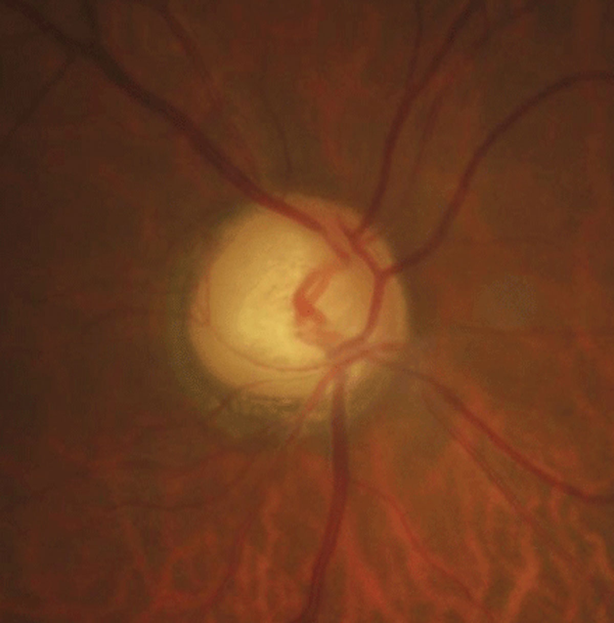
| Optic disc crescent is much more common in those with high myopia, study shows. Photo: Justin Cole, OD, and Jarett Mazzarella, OD. Click image to enlarge. |
One of the first clinically observed changes in the retina with progressing myopia is in the form of optic disc crescents, which could aid in management interventions to target those at greatest risk of progression. In this study, researchers investigated the type, dimension and appearance of optic disc crescents and how they relate to the level of myopia.
The researchers recruited 400 eyes of children and adults of a white European population ages seven to 81 with a mean spherical error of -0.50D to -14.00D. Crescent location, maximum crescent width and vertical disc diameter were measured from retinal images of right eyes only.
Six patterns of crescent were observed. Eighty-three percent of eyes had a discernible crescent. Temporal crescents were the most prevalent at 74%; however, inferior temporal crescents (16.8%) were associated with a higher degree of myopia and may be a potential marker for risk of greater myopic progression.
Only 8% of eyes in the highest category of myopia (≤-8.00D) did not have a crescent. “The high prevalence of optic disc crescents in those with low myopia (>2.00D) may be due to the inclusion of congenital crescents and the significant number of older participants in our group, whose myopia may be due to lenticular changes and the fact that age has also been associated with peripapillary changes,” the authors explained.
When excluding small crescents (<10% of the disc diameter) and re-evaluating the prevalence of crescents with age, the prevalence of crescents increased by 100% between the youngest and oldest groups. “Some of this variance may be explained if the myopia amongst those in the lower categories of myopia were still progressing, especially if they were also younger,” the authors explained.
In conclusion, higher myopia was associated with the presence of an optic disc crescent as well as larger and inferior-temporally located crescents.
“Monitoring the appearance of optic disc crescents from childhood using clinical imaging would allow study of whether crescent formation and change precedes and/or is capable of predicting the development of myopic ocular pathology,” the authors wrote.
Hill D, Heitmar R, Logan NS. Size and position of the optic disc crescent in a white European population with myopia. Ophthalmic Physiol Opt. June 20, 2022. [Epub ahead of print]. |