 |
| OCT came out on top in terms of long-term image reproducibility. Photo: Julia Reimold, OD, and Chris Wroten, OD. Click image to enlarge. |
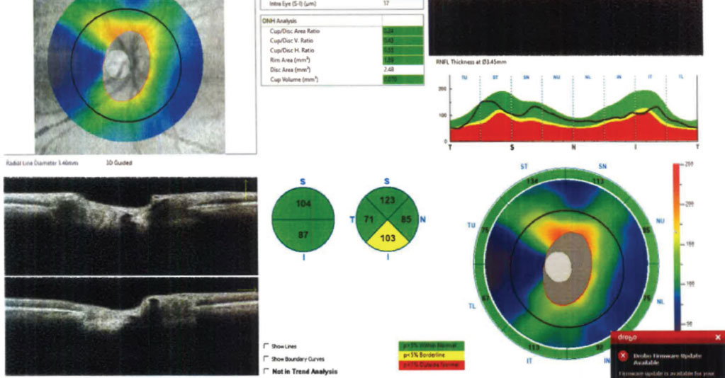
A key element to accurately identifying progression in glaucoma patients is having a set of diagnostic tests that demonstrate strong repeatability. Detection of true glaucomatous change requires the careful observation of subtle structural shifts over time, which can be made difficult by the influence of test-retest variability. Findings from a recent study suggest that when evaluating serial images for change, measuring the thickness parameters on OCT and the macula and ONH vascular parameters on OCT-A both show good long-term reproducibility, though the latter did not perform as well.
Patients in this study included those with healthy eyes (n=15), glaucoma suspect eyes (n=38) and eyes with non-progressive POAG (n=35), which was defined at the baseline visit. Each patient had at least three visits within 12 and 18 months, during which both OCT-A and OCT imaging was done on the same day using the Avanti Angiovue imaging system (Optovue). Glaucoma suspects and POAG patients all needed to be considered clinically stable to be included for longitudinal reproducibility.
The following vascular and thickness parameters were measured and assessed: macula whole image vessel density (wiVD), macula parafoveal vessel density (pfVD), ONH whole image capillary density (wiCD), ONH circumpapillary capillary density (cpCD), macular whole image ganglion cell complex (wiGCC), macular parafoveal ganglion cell complex (pfGCC) and ONH circumpapillary retinal nerve fiber layer (cpRNFL). To estimate intraclass correlation (ICC) coefficients and long-term variability, the researchers used a random effects model.
After examining the serial scans of all 88 eyes (68 subjects), they found that ICC was lower for nearly all OCT-A-measured parameters than for those on OCT, although both imaging modalities showed good long-term reproducibility and could aid in the longitudinal analysis of glaucoma patients and suspects.
“Long-term test-retest variability was between 2.29% and 2.39% for vascular parameters,” the study authors wrote. “Coefficient of variation was higher for OCT-A parameters (between 2.48% and 2.62%) compared with OCT parameters (wiGCC 0.63%, pfGCC 0.56% and cpRNFL 1.79%). Within-subject test-retest standard deviation was 1.17% and 1.22% for pfVD and cpCD, and 0.57µm and 1.22µm for pfGCC and cpRNFL, respectively.”
They found that older age and low signal strength index (SSI) were two contributing factors to decreased long-term variability of vessel densities. “The significant negative coefficients associated with age indicated that the variability of vessel density and thickness parameters increased significantly with older age,” the authors wrote. “Additionally, the significant positive associations between SSI and vessel density were found in wiVD, pfVD and wiCD, except for cpCD.”
Based on their findings, the researchers determined a cut-off value for variation that will help detect progression sooner and more accurately. They suggest that progression “may be identified when the change in vascular parameters is greater than the long-term test-retest variability of 1.9% in one year.”
Although OCT-A-measured vascular parameters scored above the threshold for acceptability, one reason why this method was still outperformed by OCT is that the image processing utilized in OCT-A may be more sensitive to image quality and more impacted by signal strength than OCT, the authors explained.
“Our results indicate that measurements over an average of 1.2 years are reproducible, supporting the use of the OCT-A as an additional tool in the long-term follow-up of glaucoma patients,” they concluded.
Nishida T, Moghimi S, Hou H, et al. Long-term reproducibility of optical coherence tomography angiography in healthy and stable glaucomatous eyes. Br J Ophthalmol. December 21, 2021. [Epub ahead of print]. |