 |
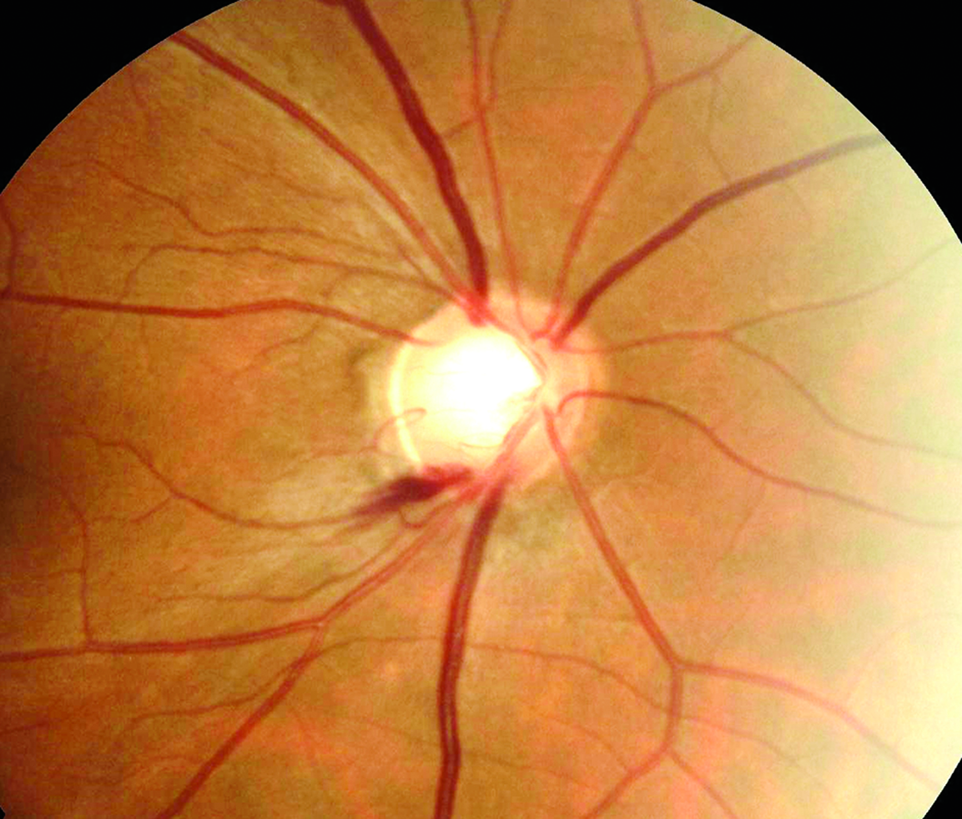
In a Chinese population, higher salt intake was linked to sooner AMD development. Photo: Sarah B. Klein, OD. Click image to enlarge. |
Detecting progression in normal-tension glaucoma patients can be challenging, as more than half of untreated patients may not display visual field progression for many years. To help identify likely progressors, researchers recently investigated the predictive value of demographics and ocular characteristics for normal-tension glaucoma progression. They found that certain reduced amplitudes on pattern electroretinography (PERG) and microvasculature dropout at baseline were significantly associated with visual field progression. Their findings are forthcoming in the Journal of Glaucoma.
The prospective study included 140 eyes with open-angle glaucoma, followed for three years. Each eye underwent at least five serial visual field tests, as well as baseline OCT-A and PERG. The researchers also obtained N35, P50 and N95 latencies and amplitudes.
The researchers reported that 76.4% of eyes had normal-tension glaucoma and 40.7% had glaucoma progression, defined by Humphrey visual field Glaucoma Progression Analysis (GPA). Mean deviation slopes were significantly different, with -0.43dB per year in progressors and 0.59dB per year in non-progressors.
Compared with glaucoma patients without progression, glaucoma patients with progression demonstrated frequent microvasculature dropout on OCT-A, isolated central scotoma, frequent disc hemorrhage and reduced baseline P50-N95 amplitude.
The researchers observed that age at diagnosis and baseline P50-N95 amplitude were significantly associated with mean deviation slope. Additionally, microvasculature dropout and baseline P50-N95 amplitude were significantly associated with visual progression on the Humphrey GPA.
“Patients with vascular features who presented with central scotoma or possess retinal ganglion cell dysfunction detected by PERG tended to show progression,” the researchers wrote in their paper. “[These patients] should be monitored closely.”
Lee M, Lopilly Park H, Kim S, et al. Predicting visual field progression by optical coherence tomography angiography and pattern electroretinography in glaucoma. J Glaucoma. July 26, 2022. [Epub ahead of print]. |