 |
|
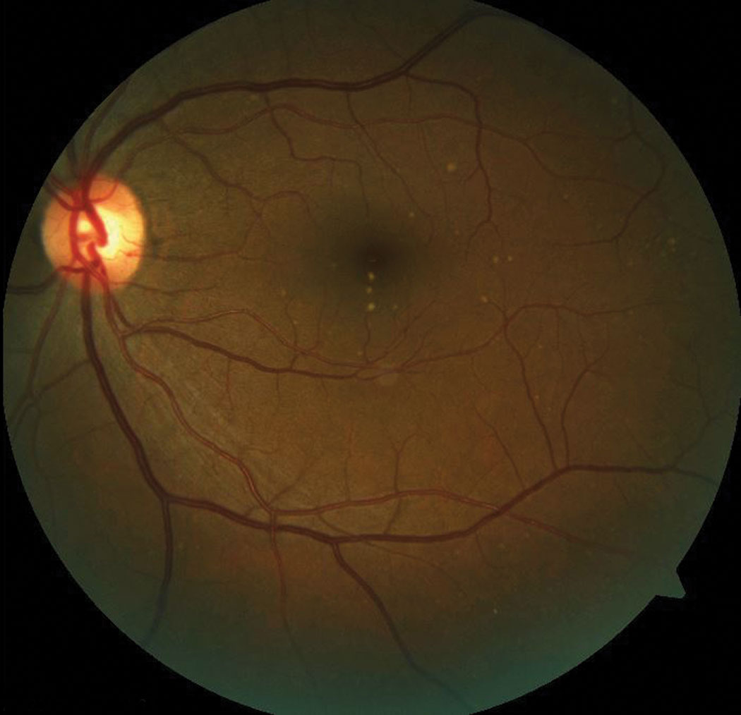
Subretinal drusenoid deposits signify photoreceptor cone loss as a higher rate than in other AMD cases. Photo: Bisant A. Labib, OD. Click image to enlarge. |
While prior research has led the way in understanding cone photoreceptor density loss in patients with age-related macular degeneration (AMD), one new study takes this facet of the disease further by including comparative measurements of patients either displaying subretinal drusenoid deposits (SDD) or not.
The prospective case series used adaptive optics scanning laser ophthalmoscopy (AOSLO) to evaluate the cone photoreceptor density in clinically unremarkable retinal regions of these patients.
Ten total eyes were studied of patients with intermediate AMD, four with SDD and three without. Measurements were taken over a period of about 40 months. Cone density was measured in 1,174 clinically unremarkable regions within the central subfield, inner and outer rings. Macular regions with an absence of AMD-associated lesions were identified through AOSLO and OCT.
Results included the finding that cone density decreased in 98.3% of all examined locations over a time period of about 40 months for those with AMD in both the SDD and drusen subcategories. Even further, decline in cone density was more rapid for those with SDD than for both the drusen and no SDD types.
At baseline, cone density was lower than normal values for both the AMD-with-drusen and AMD-with-SDD groups, with values of 31% and 42% lower than average, respectively. After about 40 months, this percentage increased significantly to 70% in the drusen group and 80% in the SDD group.
The researchers believe the reason for their findings may be related to choroidal blood flow. A deficiency of this has already been shown to play a role in AMD pathophysiology, with choriocapillaris loss seen in all AMD stages. As such, the researchers predict that reduced choroidal blood flow may affect the entire retina, resulting in retinal area cone loss without AMD lesions. Indicative of this being a possibility, eyes with SDD display thinner choroid than eyes with soft drusen and normal eyes, meaning a potential reduced metabolic capacity.
The researchers describe another possibility for reduction of cone density in the unremarkable clinical area. They specify that retinal areas without drusen or SDD can still display AMD pathology. This could look like thin accumulation of basal linear deposit or thick basal laminar deposit of the RPE-basal lamina-Bruch’s membrane complex. Seen in these areas, this pathology may be below resolving capacity in commercial imaging, but the lesions still nay contribute to cone loss.
Despite still much to be discovered following this study, the authors are hopeful that “future longitudinal studies combining confocal and non-confocal AOSLO, and autofluorescence imaging with OCT-A, may provide further insight into cellular mechanisms of disease progression in eyes with AMD by disclosing the relationship between cones, RPE cells and the choriocapillaris.” The believe their study will both provide new insight for AMD pathophysiology and foster development and assessment of photoreceptor survival therapies.
Wang X, Sadda SR, Ip M, Sarraf D, Zhang Y. In vivo longitudinal measurement of cone photoreceptor density in intermediate age-related macular degeneration. Am J Ophthalmol. 2022. [Epub ahead of print]. |