 |
|
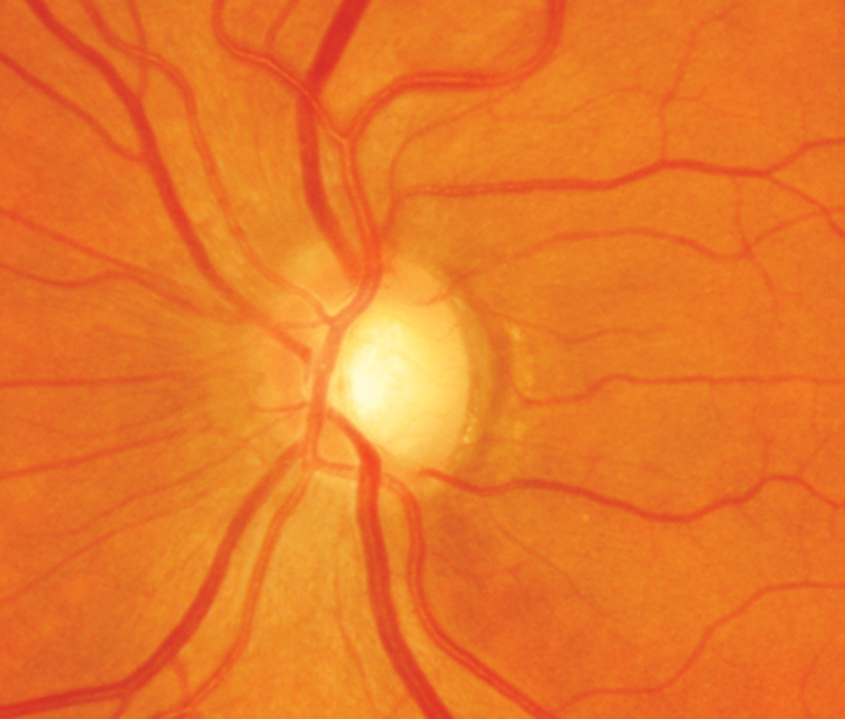
In women without glaucoma, cup-to-disc ratio was linked to lower absolute brain volume, as well as lower regional volumes in the frontal and occipital lobes. Photo: Sarah B. Klein, OD. Click image to enlarge. |
In the clinical setting, a large cup-to-disc ratio is generally associated with glaucoma patients. In addition to the normal aging process, pathologic neurodegenerative conditions of the optic nerve and the brain could lead to increased optic nerve cupping. Therefore, many believe that increased optic nerve cupping could represent physiologic aging of the optic nerve or serve as an indicator of the pathologic neurodegeneration of the optic nerve and/or the brain. A recent study determined that, in women 65 and over without glaucoma, a large cup-to-disc ratio was associated with lower relative total brain volume and absolute regional volume in the frontal and occipital lobes. Brain volume loss is associated with advanced aging and neurodegenerative conditions, including Alzheimer's and dementia-related diseases. This finding further adds to the growing evidence supporting the use of retinal imaging as a biomarker for brain health.
Retrospective data was collected from cup-to-disc ratio measurements from the Women’s Health Initiative Sight Examination study, as well as MRI-based total and regional brain volumes from the Women’s Health Initiative Memory Study MRI-1. The final analysis included 471 women without glaucoma. The mean age was 69.2, and 92.8% were Caucasian.
A large cup-to-disc ratio, defined as 0.6 or greater in either eye, was found in 7.2% of women. Controlling for total brain volume and demographic and clinical characteristics, lateral ventricle volume was 3.01cc larger, frontal lobe volume was 4.78cc lower and occipital lobe volume was 1.86cc lower for those with large ratios compared with those without.
“Our analysis suggests that there is an association between large optic nerve cupping in individuals without glaucoma and decreased brain volume,” the researchers wrote in their paper. “Enlarged optic nerve cupping in individuals without a glaucomatous diagnosis could represent a sign of optic nerve aging or be used as a surrogate measure of natural brain aging, pathologic changes and cognitive function.”
They added that subsequent studies should examine these optic disc and brain findings in larger, more racially and ethnically diverse sample sizes with other-gendered patients. Further research examining the changes in ratios in conjunction with MRI brain findings in patients with and without glaucoma is also warranted.
“It is important to note that lower global cognitive function or, specifically lower executive function could hinder a patient's abilities to perform clinical testing such as visual fields, causing them to be incorrectly labeled as glaucoma suspect or a suspect for normal-tension glaucoma,” they concluded.
Wang C, Kravets S, Sethi A, et al. An association between large optic cupping and total and regional brain volume: the Women's Health Initiative. Am J Ophthalol. January 10, 2023. [Epub ahead of print]. |
