 |
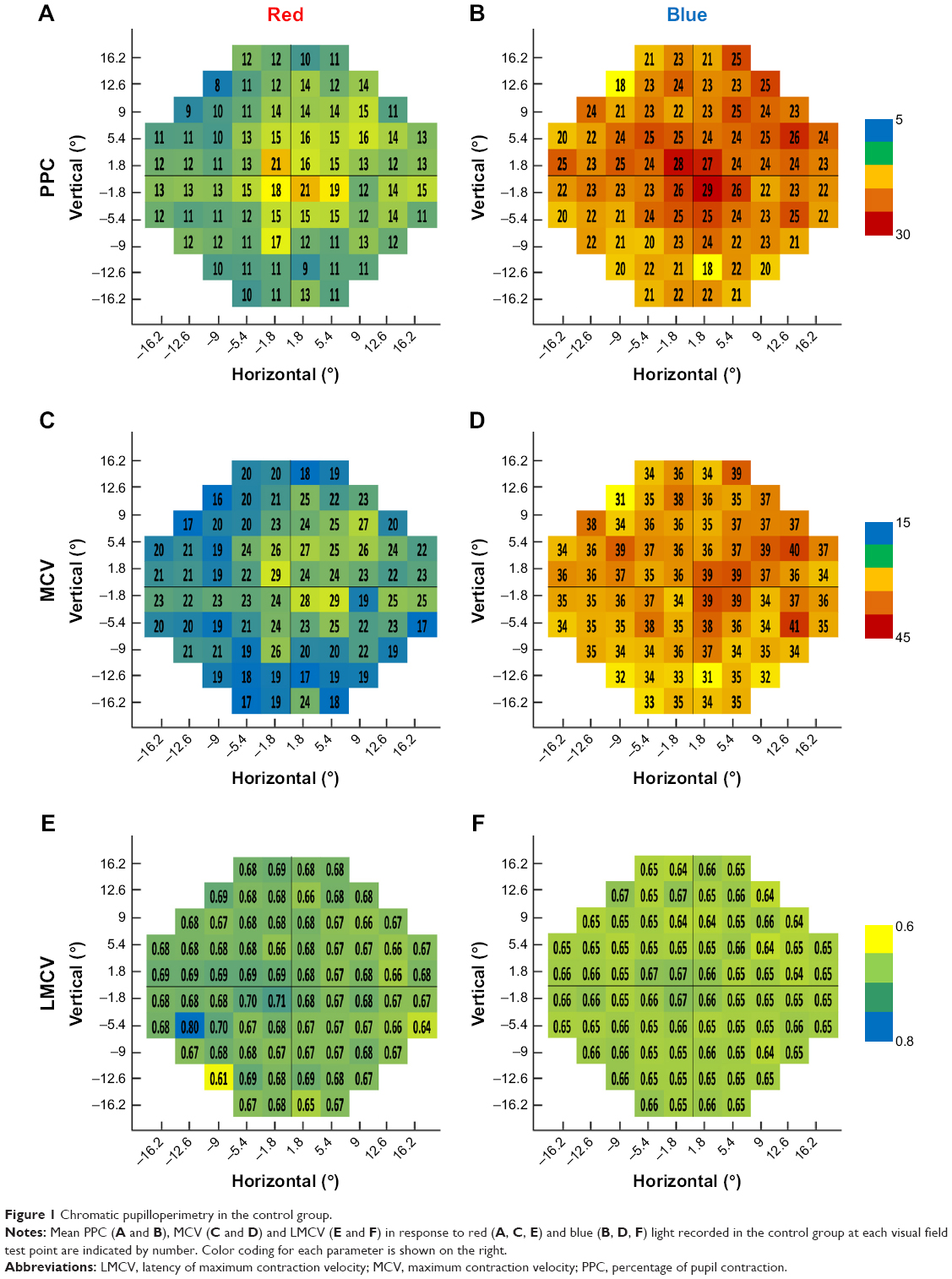
| The new technology of objective pupil light reflex testing, shown here in a study of Best vitelliform macular dystrophy, reveals new ocular manifestations of Alzheimer’s that could prove clinically useful in the future. Photo: Ben Ner D, Sher I, Hamburg A, et al. Chromatic pupilloperimetry for objective diagnosis of Best vitelliform macular dystrophy. Clin Ophthalmol. 2019;13:465-475. Click image to enlarge. |
Currently, no clinical tests or biomarkers are capable of detecting Alzheimer’s disease, although there are various signals present during a fundus exam. Given that this is the most common form of dementia worldwide, a study recently investigated whether machine learning has the potential to identify subjects at high risk of developing the condition. Its results suggested that subtle focal changes in pupil contraction latency, detected using machine learning in combination with a diagnostic technique called chromatic pupilloperimetry, may indicate a patient is at greater risk of developing Alzheimer’s even decades before clinical symptoms arise.
The study included 125 participants (45 to 71 years old) with a family history of Alzheimer’s and consequently at higher risk of disease development. In addition, 61 age-similar participants with no family history of the disease were included as controls. All subjects had normal ophthalmic assessments and retinal and optic nerve thicknesses, and no differences in cognitive function or performance or volumetric brain MRI were observed between the high-risk and control groups.
The researchers explained, “Chromatic pupilloperimetry measures the pupil light reflex for 54 small (0.43°) dim and bright red and blue light stimuli presented at a 30° visual field, thereby allowing examination of the rod-, cone- and melanopsin-mediated pupil light reflex at various retinal locations.” After conducting the test on each participant, a machine learning-based model was used to analyze the results.
The findings supported the following conclusion: “Chromatic pupilloperimetry-based machine learning models were highly discriminative in differentiating subjects with and without Alzheimer’s family history using transient pupil light reflex for focal red (primarily cone-mediated) and dim blue (primarily rod-mediated) light stimuli.” The research team also noted that features associated with transient pupil response latency achieved an area under the curve receiver operating characteristic of 0.90 for the left eye and 0.87 for the right eye.
“The test targets most discriminative of a positive Alzheimer’s family history in response to dim blue light were located in the periphery of the 24° visual field, particularly in the temporal side (nasal side of the retina),” they explained. In addition, in the right eye, the mean pupil response latency for dim blue light in the two most discriminative visual field targets in the temporal visual field was significantly shorter in individuals with a family history of the disease compared with those without. A similar trend was noted in the left eye but did not reach clinical significance.
In regard to dim red light stimuli, the researchers noted that they detected discriminative test targets throughout the retina with no specific pattern.
Although this data suggests contraction latency parameters may be predictive of high risk for Alzheimer’s disease, larger and longer studies will need to evaluate the ability of this modality to forecast actual disease development.
Lustig-Barzelay Y, Sher I, Sharvit-Ginon I, et al. Machine learning for comprehensive prediction of high risk for Alzheimer’s disease based on chromatic pupilloperimetry. Sci Rep. June 15, 2022. [Epub ahead of print]. |