 |
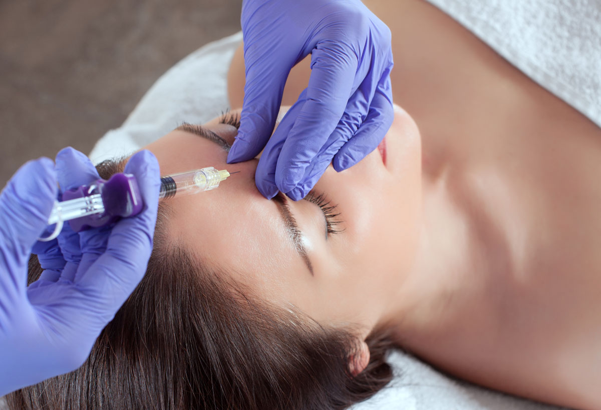
| Facial filler does not come without the occasional complication, such as CRAO, study shows. Photo: Getty Images. |
When individuals undergo cosmetic procedures such as facial filler injections, they’re not expecting to be exposed to risk an ophthalmic artery occlusion or brain infarction. They would mostly be right, as this devastating, blinding complication is quite rare. Only a few cases of iatrogenic ophthalmic artery occlusion (IOAO) and concomitant brain infarction have been reported in the literature, and the exact pathologic mechanism is still unclear.
However, researchers at Seoul National University recently reported a new case of unilateral blindness caused by IOAO and accompanied by bilateral brain infarction after a cosmetic facial filler injection. They say it’s important that patients be informed of this potential complication, even though it’s extremely rare.
In the case, a 39-year-old woman with no underlying disease presented with vision loss and ocular pain in the left eye. She also had motor weakness of her right upper and lower limbs immediately following hyaluronic acid facial filler injection into her glabella, the smooth part of the forehead above and between the eyebrows.
She underwent ophthalmic and neurologic examination, which included fundoscopic examination, slit lamp examination, fundus fluorescein angiography (FA), brain diffusion MRI and MRA. The tests revealed the following:
CRAO with choroidal ischemia
cataract, corneal edema
total ophthalmic artery occlusion
complete ptosis and total ophthalmoplegia
numerous high-signal intensity lesions in both cerebral hemispheres
multiple territorial cerebral infarction lesions involving the cerebral cortex of embolic etiology
The researchers wrote, based on these findings, that the embolism caused by filler injection was most likely the culprit behind the patient’s embolic cerebral infarction.
She underwent follow-up diffusion MRI seven days later, which revealed extensive hemorrhagic transformation. This was categorized as parenchymal hematoma, which had been noted in previous infarcted foci. Her visual field examination showed a right inferior quadrantanopia with macular sparing, which the researchers said was likely due to left occipital lobe infarction.
A review of previous literature on IOAO associated with cosmetic facial filler injection showed four cases with bilateral embolic events. Three of these were unilateral IOAO and bilateral cerebral infarction, and one was IOAO without cerebral infarction.
There are two routes through which an embolism can reach the bilateral arterial system after facial filler injection, the researchers noted—the anterior communicating artery and the cutaneous collaterals in the midline area. “In our case, IOAO occurred only on the left side,” they wrote. “According to the clinical manifestations and fundus FA findings, the embolic material caused the occlusion of the proximal part of the ophthalmic artery and could reach the internal carotid artery.”
The researchers noted that retrograde spreading of embolic materials is currently considered the most reasonable mechanism explaining IOAO. “Embolic material (typically autologous fat or hyaluronic acid) that’s accidentally injected into facial cutaneous arterioles fills the lumen of the involved vessel and may be pushed proximally against arterial flow with great injecting pressure,” they wrote. “Embolic material that reaches any branching point of the ophthalmic artery is disseminated at the branching point and is delivered to the distal portion, obstructing the distal part of the ophthalmic artery and its branches. If the amount of injected filler and injection pressure are sufficient, the embolic material can be pushed into the internal carotid artery and involve any cerebral arteries.”
In the other possible pathway, “The frontal branches of the superficial temporal artery and the supratrochlear or supraorbital artery form a network of cutaneous collateral channels,” the researchers explained. “Especially in the glabella and forehead area, numerous arterial anastomoses between the right and left cutaneous arterioles are known to exist. However, according to this hypothesis, the reason why the right cerebral infarction occurred without the right IOAO cannot be explained.” They noted that in four of the published cases, including their own, on unilateral IOAO with bilateral brain infarction, the anterior communicating artery was the most likely pathway.
“In our case, no treatment was performed for reperfusion of the ophthalmic artery because of severe clinical features, including cerebral infarction, suggesting that a large amount of filler material caused extensive obstruction of the ophthalmic artery and its branches,” they wrote. “It was suspected that the viability of the retina was lost, and cerebral infarction with mental change was a more serious problem. Hyaluronidase could be an additional treatment option. However, its effectiveness has not been proven in previous reports. Ischemic damage to the anterior segment and extraocular muscles could recover due to the relatively good collateral blood circulation and viability of tissues, which are better than those of the retina.”
The researchers concluded that clinicians should be suspicious of brain infarction, especially contralateral embolic events, in severe cases of IOAO after such procedures.
Lee J, Kim J, Woo S, et al. Unilateral blindness with bilateral brain infarction after cosmetic facial filler injection. J Neuroophthalmol 2021;41:e566-71. |