 |
|
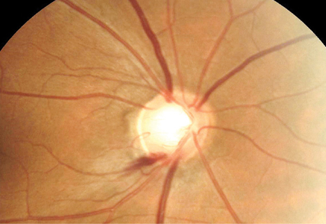
Risk factors for papillary and peripapillary hemorrhage recurrence include axial elongation, mild myopic maculopathy and glaucoma. Photo: Sarah B. Klein, OD. Click image to enlarge. |
Noting the dearth of literature and that optic nerve damage is commonly seen in eyes with pathologic myopia, researchers recently studied the prevalence and characteristics of papillary and peripapillary hemorrhages (PPHs) in eyes with pathologic myopia. They coined this new term after observing that “disc hemorrhages are located not only on the optic disc and its margins but also within the peripapillary atrophic areas surrounding the optic disc. Therefore, we have renamed disc hemorrhages as papillary and peripapillary hemorrhages.”
The researchers examined 2,171 patients (3,774 eyes) with pathologic myopia. They reported a PPH prevalence of 4.05% (88 patients [97 eyes], mean age: 66.8, mean axial length: 30.79mm). PPH recurred in 30.9% of eyes. A total of 90 eyes had single-site PPH, with the most common type and location being conus type (54.5%) and temporal side (73.3%). “Conus and periconus types of PPHs appear to be specific to pathologic myopia with a prevalence of approximately twice that of disc-related PPHs,” the researchers noted.
According to regression analysis, patchy atrophy reduced the risk of recurrences more than diffuse atrophy, and a longer axial length and potential glaucoma increased the risk. Optic nerve damage and mild myopic maculopathy were also risk factors for recurrence.
Fundus fluorescein angiography and OCT revealed that PPH developed in the area of straightened retinal arterioles in 24 eyes, at or beside the peak of a ridge in 10 eyes and in an area of compressed retinal tissue in two eyes.
“The pathogenesis of PPHs in pathologic myopia is probably different from that in glaucomatous eyes, and it may be mainly related to the mechanical tension generated by pathologic myopia-associated lesions,” the researchers concluded in their paper. “These lesions can directly or indirectly damage the vessel walls. Such mechanical forces may play a role in pathologic myopic visual field defects.”
Xiong J, Du R, Xie S, et al. Papillary and peripapillary hemorrhages in eyes with pathologic myopia. Invest Ophthalmol Vis Sci. 2022;63(12):28. |