 |
|
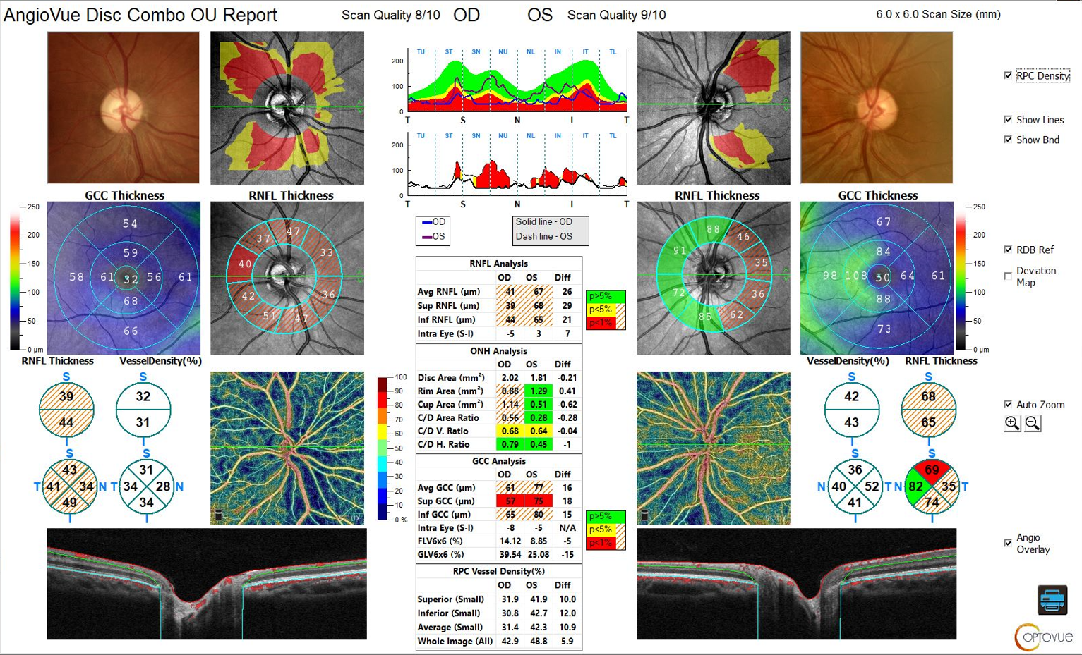
In this study, en face RNFL defects showed a greater correlation with visual field indices than did red-free RNFL defects. Photo: Visionix. Click image to enlarge. |
Evaluating retinal nerve fiber layer (RNFL) loss in patients at risk for glaucoma, such as high myopes, is crucial for earlier detection and treatment. While red-free fundus photography may only detect RNFL loss once 50% of its thickness has already been lost, OCT en face imaging can display localized RNFL defects in glaucomatous eyes and may yield clues to the detection of glaucomatous damage. In a recent study, published in the journal Eye, researchers analyzed measurements from the two imaging modalities to investigate the correlation and agreement of RNFL defects obtained from both, as well as compared them for the strength of the structure-function association. They found that en face RNFL defects showed a stronger correlation with visual field loss severity than red-free RNFL defects in eyes with and without myopia.
Included in the study were 256 eyes of 256 patients with localized RNFL defects on red-free fundus photography, as well as a subgroup of 81 highly myopic eyes (≤-6.00D). The researchers observed that in 91% of eyes, the angular width of en face RNFL defects was smaller than that of red-free RNFL defects. Additionally, the association of en face RNFL defects with mean deviation and pattern standard deviation was stronger than that of red-free RNFL defects, especially in highly myopic eyes.
In their paper, the researchers elaborated on their findings. “The angular widths between the red-free and en face RNFL defects showed a large discrepancy,” they noted, adding that this observation contrasts prior studies. “En face RNFL defects reflected visual function better than did red-free RNFL defects. Furthermore, this structure-function association of en face RNFL defects was much stronger in highly myopic eyes.”
The team went on to conclude that “angular measurement of RNFL defects by OCT en face imaging is significantly associated with the severity of visual field loss.” Based on this data, they suggest that OCT en face imaging could help improve the accuracy of glaucoma detection and monitoring. Further research on the structure-function association of glaucoma may be useful to inform disease severity grading, as well as to deepen our understanding of the natural history of the complex condition.
Bak E, Choi HJ. Structure-function relationship in glaucoma: optical coherence tomography en face imaging vs. red-free fundus photography. Eye. February 23, 2023. [Epub ahead of print]. |
