 |
|
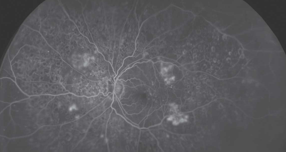
Ultra-widefield imaging showed an association between peripheral lesions and nonperfusion with DR disease worsening. Photo: Optos. Click image to enlarge. |
Imaging advances in ultra-widefield (UWF) technology allow broader views of the retinal periphery as well as the ability to capture posterior and peripheral retinal nonperfusion on UWF fluorescein angiography (FA). A new study from the DRCR Retina Network (called Protocol AA) evaluated whether UWF fundus photography and FA imaging can improve the ability to predict rates of DR progression beyond what is provided by the current standard Early Treatment Diabetic Retinopathy Study Diabetic Retinopathy Severity Scale (ETDRS DRSS). The four-year longitudinal study, as evidenced in published articles in JAMA Ophthalmology, demonstrated that both greater baseline retinal nonperfusion and predominantly peripheral DR lesions on UWF-FA are associated with higher risk of disease worsening, even after adjusting for baseline DRSS score and known systemic risk.1,2
The cohort study was a prospective, multicenter, longitudinal observational study conducted at 37 US and Canadian sites. At baseline and annually through four years, 200° UWF color images were obtained and graded for DRSS at a reading center. Disease worsening was defined as a worsening of two steps or more on the DRSS or receipt of DR treatment. The peripheral lesion study analyzed 544 eyes with nonproliferative DR (50% female participants; median age 62; 68% Caucasian). Predominantly peripheral lesions were defined as DR lesions with a greater extent outside vs. inside the seven standard ETDRS fields.1
Although no association was identified with color lesions, the presence of predominantly peripheral lesions on FA was associated with a significantly greater risk of ETDRS DRSS worsening or treatment over four years independent of DRSS score. Eyes with FA lesions had a 1.7-fold greater risk of nonproliferative DR worsening over four years. The four-year disease worsening rates were 45% for eyes with baseline mild, 40% for moderate, 26% for moderately severe and 43% for severe nonproliferative DR.1
“These results suggest that use of UWF-FA to evaluate retinas peripheral to standard ETDRS fields may improve the ability to predict disease worsening in nonproliferative DR eyes,” the researchers wrote in their paper.1
The nonperfusion study examined 508 eyes of the protocol’s cohort. After adjusting for baseline DRSS, the risk of disease worsening over four years was higher in eyes with greater overall nonperfusion index and within the posterior and midperiphery.2
“Although greater extent and severity of retinal nonperfusion was associated with the presence of FA predominantly peripheral lesions, both nonperfusion and these lesions are independently associated with DRSS worsening or treatment even after adjusting for baseline DRSS score, diabetes duration and HbA1c value,” the researchers wrote in their paper.
“These associations between disease worsening and retinal nonperfusion and FA predominantly peripheral lesions support the increased use of UWF-FA to complement color fundus photography in future efforts for DR prognosis, clinical care and research,” they concluded.2
1. Marcus DM, Silva PS, Liu D, et al. Association of predominantly peripheral lesions on ultra-widefield imaging and the risk of diabetic retinopathy worsening over time. JAMA Ophthalmol. August 18, 2022. [Epub ahead of print]. 2. Silva PS, Marcus DM, Liu D, et al. Association of ultra-widefield fluorescein angiography-identified retinal nonperfusion and the risk of diabetic retinopathy worsening over time. JAMA Ophthalmol. August 18, 2022. [Epub ahead of print]. |
