 |
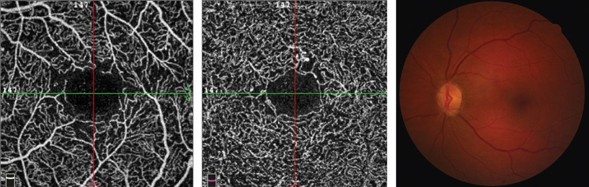
| Differences in FAZ size at birth may arise from incomplete cleavage of the inner retinal layers. Photo: Julie Rodman, OD. Click image to enlarge. |
Differentiating a normal retina from one that falls outside the norm relies on the availability of information and data that characterize how a healthy retina should look and function. A recent study observed a group of children with healthy eyes and no comorbidities to help define a spectrum of normal findings. The data revealed a correlation between foveal thickness, fovea-to-macula thickness ratio and foveal microvasculature measurements. In addition, the researchers found fovea-to-macula thickness ratio to be a reliable and quantifiable way to assess the structural development of the fovea.
The observational, cross-sectional study included 86 children between ages eight and 17 born at full-term with no eye or systemic disease. OCT-A scans (3x3mm) were done on each child, as was visual acuity testing. Imaging software collected measurements of vessel density and perfusion, while structural measures including foveal and macular thicknesses were performed manually by the researchers.
The following average measurements were recorded in the cohort: BCVA -0.10 logMAR, refractive error 0.59D, axial length 23.1mm and area of the foveal avascular zone 0.20mm2. The median fovea-to-macula thickness ratio was 0.63, mean central vessel density was 12.42mm and mean central perfusion was 38.7%.
In addition, the researchers reported, “Area of the foveal avascular zone was correlated with central vessel density, perfusion, foveal thickness and fovea-to-macula thickness ratio. Central vessel density was correlated with foveal thickness and fovea-to-macula thickness ratio.” Lastly, they observed, “Central perfusion was correlated with foveal thickness and fovea-to-macula thickness ratio.”
They noted, “The most important finding was the relationship between the area of the foveal avascular zone and the fovea-to-macula thickness ratio. Thicker foveae were associated with increased vascular density and perfusion.” Children with shallower pits, and therefore higher fovea-to-macula thickness ratios, had a smaller area of the foveal avascular zone, on average. The researchers suggested these parameters may be associated with incomplete foveal development, though the mechanisms and implications of this association remain unknown.
“Clinicians need to be aware of this variability of the fovea in both children and adults when making management decisions,” the researchers concluded.
Demir P, Hovsepian N, Pagels P, et al. All retinas are not created equal: fovea-to-macula thickness ratio and foveal microvasculature in healthy young children. Ophthalmic Physiol Opt. January 12, 2022. [Epub ahead of print]. |