 |
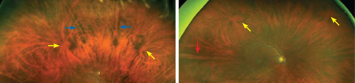
Myopic patients had about 10% increased sensitivity for peripheral lesion detection using ultra-widefield imaging. Photo: Mohammad Rafieetary, OD. Click image to enlarge. |
Ultra-widefield imaging (UWFI) has been used in clinical practice for assessing and photo-documenting vitreoretinal pathologies, with an advantage of visualizing the peripheral area, but it isn’t yet clear if mydriasis can increase the sensitivity of detecting peripheral retinal lesions through an eye-steering UWFI technique, which is what researchers sought to determine in a recent study.
A total of 220 eyes of 110 myopic patients with peripheral retinal lesions in at least one eye were recruited. Non-mydriatic and mydriatic UWFI images were taken centrally and in upper, lower, nasal and temporal gazes with the Optomap UWFI. Sensitivity of detecting peripheral retinal lesions under different UWFI settings was then compared.
Of the study eyes, 64% had peripheral retinal lesions. Mydriasis helped increase the sensitivity in standard gaze by around 10% for peripheral retinal lesions, improving retinal hole and degeneration detection as well, which is consistent with results from a previous study. The eye-steering technique significantly improved the sensitivity of detecting peripheral lesions by more than 30%, not only with the non-mydriatic method but also under mydriatic circumstance.
Lesions of the superior and inferior quadrants benefited more from the eye-steering technique, which overcame the disadvantage of the UWFI design that used an elliptical mirror, the authors noted.
“A detectable lesion under UWFI could not be 100% detected in the corresponding gaze, even under the mydriatic condition. One reason is that the detected retinal area in four corresponding gazes was not strictly the same as the divided four quadrants,” the authors explained. “Another reason is, as stated above, the disparity of captured retinal area under UWFI.”
Peripheral lesions that needed prophylactic treatment did not have a difference in detection compared with those needing no treatment under all four settings of UWFI.
“Neither axial length nor spherical equivalence influenced the sensitivity of detecting peripheral retinal lesions in each UWFI setting, which actually confirmed the clinical experience,” the authors explained. “And previous studies have already proved that high myopia and long axial length had more impact on the sharpness of peripheral retina rather than the visualized retinal area.”
Li M, Yang D, Shen Y, et al. Application of mydriasis and eye steering in ultrawide field imaging for detecting peripheral retinal lesions in myopic patients. Br J Ophthalmol. March 3, 2022. [Epub ahead of print]. |