 |
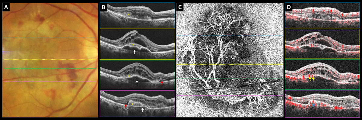
| In type 2 macular neovascularization cases like this one, decreased retinal sensitivity likely results from new vessel growth between the neurosensory retina and RPE, study finds. White arrows point to PED, yellow asterisk shows subretinal neovascular membrane, red arrows highlight subretinal fluid, yellow arrows indicate subretinal feeder vessels and blue arrow points to subretinal neovascular membrane. Photo: Carolyn Majcher, OD. Click image to enlarge. |
Researchers recently evaluated diagnostic factors associated with exudative age-related macular degeneration (AMD) to pinpoint more effective markers. The prospective design included 24 eyes from 24 patients in a two-year study of PRN bevacizumab treatment for wet AMD. Over this period, visual acuity, fluorescein and indocyanine green angiographies, autofluorescence images, microperimetries and OCTs were analyzed.
Microperimetry results were aligned with the OCTs, angiographies and autofluorescence images. Each stimulus site was measured for neuroretinal thickness, retinal pigment epithelial (RPE) elevation, neuroepithelial detachment, subretinal tissue and cystic intraretinal fluid, and indocyanine green plaque, hemorrhage and RPE atrophy were identified.
The study researchers found that microperimetric retinal sensitivity increased during the first year but remained constant during the second. At one year, baseline lesion components that most strongly aligned with predicting deteriorated sensitivity were RPE atrophy, the area of type 2 macular neovascularization, intraretinal cysts, hemorrhage, type 1 neovascularization and retinal thickening greater than 350μm. Only minimal effects were seen with neuroepithelial detachment and RPE elevation. The predictive values of the baseline lesion components remained mostly unchanged after the two years.
The authors pointed out, in their paper for Acta Ophthalmologica, that the decreased focal retinal sensitivity seen in these components was associated with decreased visual acuity in prior studies. However, they specified that visual acuity does not give great insight into tissue changes resulting in this deteriorated function.
This study’s use of microperimetry and retinal OCT allowed the authors to gather information about sensitivity and structure-specific loci of the macula, which is different from prior studies. Earlier studies have used microperimetry values superimposed on color fundus photographs and fluorescein angiograms or aligned a sensitivity map manually onto OCT images. Results from these studies showed the least improvement occurred in loci with intraretinal cysts, while best prognosis was related to morphology with subretinal fluid and serous pigment epithelial detachment.
Earlier research also having found noticeable retinal sensitivity improvement after one year of anti-VEGF treatment but staying stagnant at two years corroborates this study’s findings, the authors pointed out.
Decline in retinal sensitivity seen with atrophy, the authors note, is caused by insufficient photoreceptors, while hemorrhaging can result in optical hindrance to viewing. With type 2 macular neovascularization, decreased sensitivity is likely resultant of new vessels between the neurosensory retina and RPE. The correlation between cysts and reduced sensitivity identifies cysts as a marker of retinal dysfunction as well.
The researchers concluded, “It appears that quite similar sets of lesion components are associated with baseline, one- and two-year retinal sensitivity losses and their prediction. Thus, a decline in visual activity during a longer follow-up is probably due to an enlargement of the same lesion components rather than the emergence of new ones.” Knowledge of these underlying changes allows for better treatment to follow suit.
Bygglin H, Immonen I, Luoma A, Hautamäki A. Exudative age-related macular degeneration lesion components predicting microperimetric retinal sensitivity during anti-vascular endothelial growth factor treatment. Acta Ophthalmol. May 25, 2023. [Epub ahead of print]. |