 |
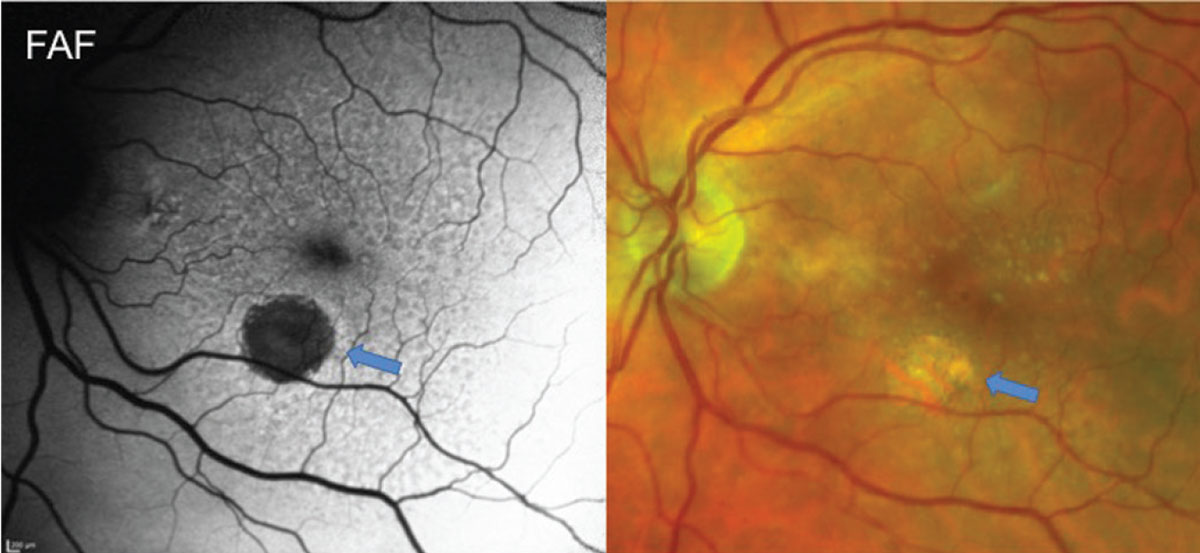
| This study determined that the presence of reticular pseudodrusen carried a higher risk for GA than nAMD. Photo: Jessica Haynes, OD. Click image to enlarge. |
A post-hoc analysis of two clinical trial cohorts—Age-Related Eye Disease Study (AREDS) and AREDS2—recently found that reticular pseudodrusen serves as an anatomical risk factor for progression to late-stage age-related macular degeneration (AMD).
This study included eyes with no late AMD at baseline in AREDS (n=6,959 eyes, 3,780 participants) and AREDS2 (n=3,355 eyes, 2,056 participants). The main outcome measures included progression to late AMD, geographic atrophy and neovascular AMD.
The researchers graded color fundus photographs from annual study visits for soft drusen, pigmentary abnormalities and late AMD. The presence of reticular pseudodrusen was determined by grading of fundus autofluorescence images in AREDS2 and deep learning grading of color fundus photographs in AREDS. The team performed proportional hazard regression analyses, considering AREDS AMD severity scale and reticular pseudodrusen at the same time.
In AREDS, the study authors reported a correlation between the presence of reticular pseudodrusen and a higher risk of progression to late-stage disease. However, they observed significant differences in the associated risk at simplified severity scale levels. The nine-step scale also showed a higher risk of progression when reticular pseudodrusen is present.
The investigators also observed a significant increase in the risk of progression among the AREDS2 study population when reticular pseudodrusen presence was defined directly by reading center grading of fundus autofluorescence images. In both cohorts, the study authors found that the presence of reticular pseudodrusen carried a higher risk for geographic atrophy than neovascular AMD.
“Reticular pseudodrusen represents an important anatomical risk factor for progression to late AMD, particularly geographic atrophy. However, the added risk associated with reticular pseudodrusen varies markedly by severity level,” the study authors noted. “It carries highly increased risk at lower/moderate levels and less increased risk at higher levels.”
For an accurate understating of progression risk and subtype predictions, they concluded that reticular pseudodrusen status should be considered in combination with traditional features. Moving forward, it should also be included in updated AMD classification systems and risk calculators as well as clinical trials.
Agrón E, Domalpally A, Cukras CA, et al. Reticular pseudodrusen: the third macular risk feature for progression to late age-related macular degeneration. Ophthalmology. May 31, 2022. [Epub ahead of print]. |