 |
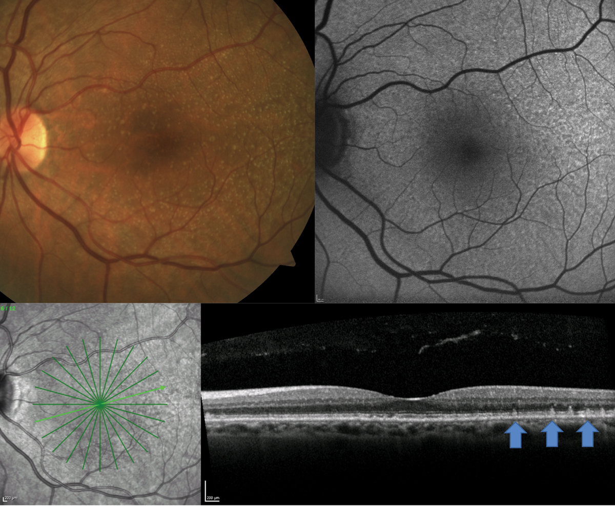
| Eyes with reticular pseudodrusen had significant retinal structural and vascular changes compared with intermediate dry AMD eyes. Photo: Jessica Haynes, OD. Click image to enlarge. |
Drusen is one of the hallmarks of age-related macular degeneration (AMD) and a key feature used in disease classification. One particular type called reticular pseudodrusen (RPD)—yellow, interlacing structures in the outer macula—was recently examined in a study published this month in Ophthalmic Research. RPD are thought to be biochemically different from soft drusen, with a different lipid composition and a higher concentration of unesterified cholesterol. Employing improvements in multimodal imaging and OCT-A, the researchers studied RPD and its effects on the retina, hypothesizing that RPD might be part of a modified disease process associated with drusen alone. They found that patients with RPD had inner retinal structural thinning and vascular changes—more so than patients with only intermediate dry AMD.
The researchers retrospectively analyzed 25 eyes of 17 patients with RPD, 20 eyes of 15 patients with intermediate dry AMD and 14 eyes of nine patients with both. Each patient had undergone imaging and measurement of the central 3mm retinal thickness, which was subdivided into nine ETDRS sectors.
Based on retinal thickness analyses, the team found that patients with both intermediate dry AMD and RPD had significantly thinner superior inner and superior outer maculae than patients with only intermediate dry AMD. For RPD eyes, the superior inner and superior outer retinal pigment epithelium, outer plexiform layer and inner nuclear layer were also thinner than in eyes with only intermediate dry AMD. These eyes also had significantly reduced macular deep capillary plexus vessel density compared with intermediate dry AMD.
“Previous authors have found retinal thinning accompanied by retinal vascular loss in AMD patients with RPD,” the authors wrote in their paper. “The inner retinal layers are supplied by the superior capillary plexus and deep capillary plexus, and a greater portion of the outer plexiform layer receives nourishment from the choriocapillaris indirectly. Interestingly, in the present study, deep capillary plexus vessel density was significantly lower among patients with RPD than those with intermediate dry AMD. This may be due to inner retinal layers showing the highest sensitivity to hypoxic challenges, whereas the outer retina is more resistant to hypoxic stress.”
According to the authors, several questions remain. They wrote that it’s still unclear why inner retinal vascularity is reduced from RPD, which is found in the outer retina, and that they’re also uncertain whether retinal thinning or reduced vascularity comes first. “RPD is known to affect the outer retina, and there may be some transsynaptic degeneration which in turn leads to reduced vascularity,” they suggested. “Although the main focus of our study was on inner retinal changes, our study also found that the retinal pigment epithelium was significantly thinner among eyes with RPD than in those diagnosed with intermediate dry AMD.”
The researchers concluded that their observations “regarding thinning and vessel density loss should be investigated further.”
Kalaw FGP, Alex V, Walker E, et al. Inner retinal thickness and vasculature in patients with reticular pseudodrusen. Ophthalmic Res. June 2, 2023. [Epub ahead of print]. |