 Q: I work with an excellent cataract surgeon who is an hour away, and I have a patient with an IOP of 40mm Hg in for the one-day visit. The surgeon asked me to “burp” the wound. How do I do it?
A: “Burping” the wound is definitely something that optometrists can do, says Howell Findley, O.D., who typically burps one or two eyes a month at Commonwealth Eye Care, a comanagement and ocular surgery center in Lexington, Ky.
The hardest part is having the confidence to do it the first time, Dr. Findley says. “It’s a learnable skill—but like anything else, if you’re not comfortable with it, don’t do it.”
Burping a wound is nothing more than putting pressure against the paracentesis incision, which opens it up to release a build-up of aqueous.
Why has the aqueous built up and caused the pressure to rise? Because there’s likely still viscoelastic remaining from the cataract surgery, which has clogged up the trabecular meshwork and caused a blockage, with a subsequent spike in pressure.
This obstruction will clear eventually; but until it does, the aqueous has to go somewhere—and the simplest exit is through one of the surgical incisions. That’s where the burp comes in. Here’s how it’s done, Dr. Findley explains:
1. Locate the paracentesis. If the surgeon has made a clear corneal incision, then this side-port incision is usually about two clock hours away from the main incision. “So if the main incision in the left eye is at 12 o’clock, then the paracentesis is going to be at about 2 o’clock,” Dr. Findley says. “If the incision in the right eye is at 9 o’clock, then the paracentesis wound is going to be somewhere around 10 to 11.”
2. Choose the right instrument. Once you’ve located the paracentesis, you need the right tool for the job: Avoid a sharp one, if you can. “Some doctors use a Beaver blade or a bent needle,” Dr. Findley says. “But the potential danger with one of those is that, if the patient moves his eye, you could poke through the sclera or elongate the paracentesis wound. So, I prefer to use a blunt-tipped probe.”
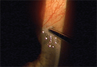
1. How do you do a “burp”? First, place a blunt-tipped probe just posterior to the paracentesis wound.
2. Apply pressure on the posterior side of the paracentesis to allow aqueous fluid to escape, which lowers IOP.
3. A half a second of pressure is usually enough. Here, a small gush of aqueous flows onto lower eyelid after the burp.
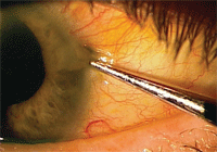
4. Pressure from the
instrument may leave a temporary indentation on the sclera. It’s
unsightly, but not a problem. Now measure IOP to ensure it went down.
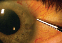
3. Apply the instrument just outside of the wound. Place the tip of the instrument as close as you can to the outside (limbal) edge of the paracentesis wound. Then push straight in.
Usually it’s easy, but be prepared for some resistance, Dr. Findley says. Because it’s a self-sealing wound, the intraocular pressure wants to keep it closed. So if the pressure is higher (60mm Hg or more), the wound might be harder to open.
4. Release the pressure. As soon as you get the wound open, you should see a little gush of clear aqueous fluid emerge. You’ve done it! Now quickly remove the instrument, which should close the wound. Burping the wound for a half-second in a patient with an IOP of 40mm Hg should usually do the trick, or perhaps a full second for a patient with an IOP of 50mm Hg or 60mm Hg, Dr. Findley says.
5. Check the pressure. Measure the patient’s IOP to see how far it has come down. “You can immediately take a pressure from 40mm Hg or 50mm Hg down to the teens, which is where you want it,” he says. “While you’re at it, make sure that the pupil is still round, the IOL is centered, and the anterior chamber is formed.”
A half hour later, re-check the patient’s IOP to make sure it has stayed down. Have the patient return the next day to measure it once more. If the IOP has risen, either at the half-hour point or the next day, burp the wound again.
6. Add medical support. If the patient has no allergy to sulfa, start acetazolamide, 250mg q.i.d. for a day or two, just to help keep the pressure down.
In addition, double the dose of the patient’s antibiotic for a day or two. “In 25 years, I’ve never seen a patient get an infection from this,” Dr. Findley says. “But I still feel better if we kick the antibiotics up a notch.”
Q: What risks are involved? Is this even within our scope of practice?
A: The main risk is in not doing it, Dr. Findley says. If the patient already has glaucoma and gets a pressure spike, you need to bring that pressure down soon or there could be permanent optic nerve damage.
In that regard, a general threshold is about 40mm Hg or more for burping the wound. But a patient with pressure in the 20s or 30s can probably get by with medical management and more frequent IOP checks.
Another possible risk in a “floppy iris” patient on Flomax (tamsulosin, Boehringer Ingelheim) is the iris being sucked up into the wound. “Instead of burping it, give the patient acetazolamide and call the surgeon,” Dr. Findley says.
Lastly, “burping a wound should certainly be within our scope of practice because we’re not cutting or inserting anything into the eye,” he says.
However, just to cover your bases, don’t do it on your own without communicating with the surgeon first, or establishing standing orders or protocols when the situation occurs.
