Level Up Your TechOptometrists today have more tools than ever at their disposal to help detect, treat and manage disease. To help you keep up with the constant innovation, the September issue of Review of Optometry—our 46th annual technology report—gives readers a rundown on what's new in FAF imaging, visual field testing, ultra-widefield imaging, low vision and more. |
Optometrists are often the first providers to see patients experiencing vision loss, whether new, progressive or long-standing. Although it is the duty of the provider to diagnose and manage the underlying cause of the worsening vision, we cannot overlook how irreversible vision loss impacts quality of life for our patients. It is crucial as eyecare professionals and experts in our field to fully understand the needs of this patient population beyond the exam chair, which may be as simple as having an in-office conversation.
It is also important to remember that there is no “acuity cutoff” necessary for a low vision rehabilitation referral. Any patient with a history of ocular disease whose activities of daily living are not being met should be promptly referred to a low vision rehabilitation (LVR) specialist. LVR aims to use resources and tools in order to help patients adapt to lifestyle changes as a result of vision loss. In this article, not only will the importance of LVR referral be highlighted, but also the need for primary eyecare practitioners to become aware of possible tools and resources so their affected patients may benefit from these services, especially if an LVR provider is not in their area.
Where to Start?
It is the duty of every eye doctor to promptly and accurately refer patients to specialists when needed, and low vision rehabilitation is no exception. At a minimum, our profession must be familiar with state agencies and organizations for the visually impaired and how to register a patient when necessary as legally blind, which will allow patients to access available resources. These resources can often be found online by searching your state’s government websites.
For example, at the time this article was written, to register in Massachusetts for legal blindness, an online form can be completed and submitted to the Massachusetts Commission for the Blind. In many states, eye doctors have a legal duty to register their patients promptly in order to provide them with resources they have a right to receive. Following registration, patients are usually connected with a case manager, generally a social worker, who will act as their liaison. This may vary state to state, but it is crucial to connect patients with accurate resources, and to do so requires the first step of registration.
Even when a patient does not qualify for legal blindness but does have vision loss, it is imperative that eye doctors are aware of alternative options to help meet the needs of their patients. Such important resources include grief counseling and support groups. This is incredibly vital not to overlook, as many patients struggle with progressive or sudden vision loss. There are groups specific to veterans through local Veterans Hospitals that patients may get involved with, but many local programs exist as well. Reaching out to your state’s commission for blindness or foundation for blindness is a great way to learn about these groups and how to connect them with your patients.
When any patient has experienced impact to their daily activities because of vision changes, thorough consideration should also be given to an LVR referral. Having a knowledgeable conversation with patients about the potential advantages these referrals offer is crucial to determine if the patient is motivated and will actually benefit from a referral. Searching online and asking others in the field are some ways to find an LVR provider, but reaching out to the nearest optometry school is another productive option that can be overlooked. Additionally, many large ophthalmology programs have optometrists (and less frequently ophthalmologists) practicing LVR that are affiliated with their hospitals.
Remember, LVR examinations are goal-specific. Providers listen carefully to the needs of their patients and identify areas in which low vision technology and resources may be of adequate assistance. To accurately identify these resources and complete a comprehensive LVR examination, understanding the connection between the disease process and a patient’s functional vision loss is key. Although not all-inclusive, this article provides a brief overview of common causes of vision loss and the impact these conditions may commonly play in quality of life. Also discussed are recommendations an LVR provider may consider in an effort to encourage primary eyecare providers to begin the conversation about low vision and its resources to further understand why LVR referral is key.
Central Scotomas and Metamorphopsia
A central scotoma is a blind spot or area of decreased vision impacting central vision, while metamorphopsia is described as vision distortion. Both are often experienced by patients suffering from conditions like age-related macular degeneration and Stargardt’s disease.
Many patients with central scotomas and metamorphopsia may have trouble reading, especially continuous text, as the distortion or blind spot may cause letters to appear jumbled or missing. Central scotomas may also reduce the amount of saccadic eye movements during reading, greatly impairing reading speed and efficiency.1 This form of vision loss may also significantly impact one’s ability to drive confidently or recognize faces, as central vision is impaired or missing entirely.
 |
|
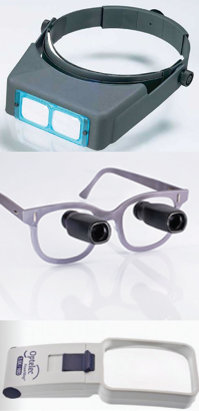
Fig. 1. Top: Optivisor with light for intermediate tasks. Middle: Spectacle-mounted telescope for distance enhancement. Bottom: Optelec illuminated handheld magnifier for spot reading. Click image to enlarge. |
Patients with central scotomas are often very successful with eccentric viewing, magnification and additional goal-specific strategies. Eccentric viewing is a method of vision training that works to move the viewed image into the patient’s preferred retinal locus (PRL).2,3 Patients may learn to use their PRL to improve visual clarity by using a less damaged or healthy part of their retina.4,5
This training can be completed in-office by the OD, but is often completed by a referred specialist, such as a low vision-trained occupational therapist, certified low vision therapist or vision rehabilitation therapist. Eye doctors should be familiar with vision rehabilitation specialists like these in their area. Aside from a quick Google search or word-of-mouth recommendations, using the following website can direct your efforts: www.acvrep.org/index.
As for task-specific goals, this is where referral to a low vision rehabilitation provider may be necessary. Most eye doctors have some (albeit varying) level of experience with LVR. As the field has grown extensively beyond high-powered adds and magnifiers, a full LVR exam is difficult to complete in a primary care setting.
During the LVR examination, specialists will thoroughly review the patient’s needs and determine which potential devices and resources they may benefit from. These may range from optical devices such as handheld magnifiers, stand magnifiers, microscopes and telescopes to electronic devices and assistive technology on phones and computers, either built into the software or through third-party applications. Electronic devices to consider include desktop magnifiers, electronic magnifiers and head-mounted devices. Many of these allow for adjustable magnification, reverse polarity and audibility.6
Peripheral Vision Loss
This type of loss constitutes a gradual reduction of vision moving from peripheral to central regions. It is often experienced by patients suffering from conditions such as glaucoma and retinitis pigmentosa.
Many patients with peripheral vision loss may have difficulty with mobility and express this in the form of bumping into objects or missing objects as they come towards them.7 This may result in an increased risk of falling and lack of confidence when traveling independently. Although not all states have a visual field requirement for legal driving, this difficulty with peripheral vision loss may make it difficult or impossible to drive. Additionally, smaller fields of vision may hinder reading abilities, as the patient may find it difficult to localize words on a page or it may be harder to read efficiently with fewer words in their field of view.8
 |
|
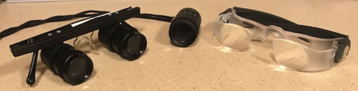
Fig. 2. Left to right: Binocular telescope, monocular telescope and MaxTV glasses for distance enhancement. Click image to enlarge. |
Tools such as reverse telescopes prove successful with patients experiencing peripheral vision loss. The use of a monocular telescope in the reverse direction minifies objects and expands the patient’s field of view, therefore allowing a greater number of smaller images within the field of vision.9 This can be especially beneficial for mobility purposes. Following evaluation and recommendation from an LVR provider of which type of reverse telescope to use, a qualified rehabilitation specialist is useful for possible in-home training with the device.
Another important resource for referrals is a certified orientation and mobility specialist (COMS). These specialists work to train patients with vision loss so they are able to travel and freely move about their home, work and public environments.10 This is often the best way to provide patients with white cane training and broaden their independence regarding mobility concerns.
 |
|
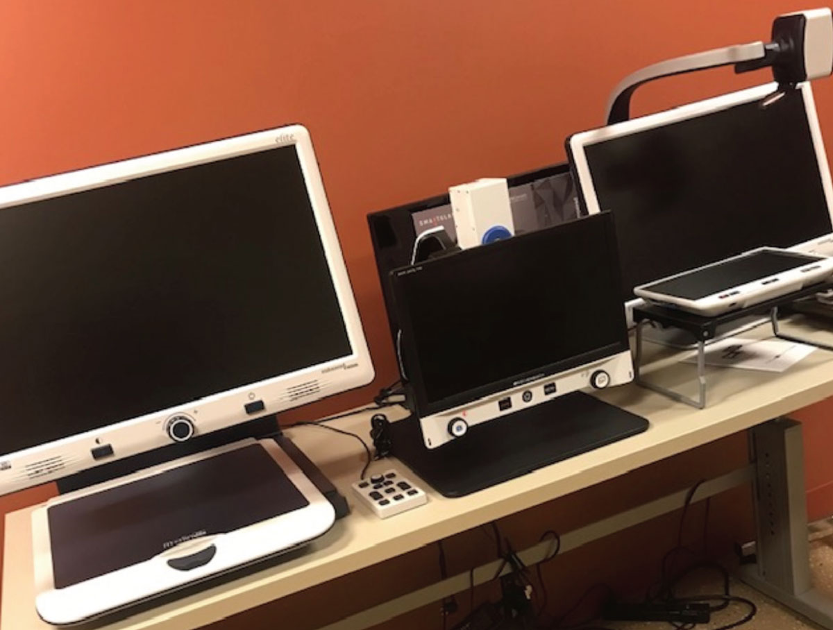
Fig. 3. Left to right: Distance-enhancement interfaces corresponding to the aforementioned binocular telescope, monocular telescope and Max TV glasses of Fig. 2, respectively. Click image to enlarge. |
Hemianopsia and Quadranopsia
Vision loss of this type is the loss of half or a quarter of the patient’s vision bilaterally. This form of vision loss is often experienced by patients suffering from conditions such as trauma and stroke.
Like patients with full peripheral vision loss, patients with partial loss also have trouble with mobility, object localization, driving and reading.11 Especially in cases of right-sided loss, the patient may be reading into their scotoma and have trouble discerning where the end of the page is located.
Outside of an LVR consultation, referral to a COMS and any other applicable rehabilitation specialist (as determined by the patient’s goals) is best practice for many patients with hemianopsia field loss. A prism evaluation may be extremely useful for these patients. Use of prisms, including but not limited to Peli prism, yoked prism and Fresnel prism may require additional training that can be provided either in-office or by these specialists.12,13
Contrast Sensitivity and Glare
These factors are arguably two of the most overlooked aspects of vision reduction. A thorough evaluation may be completed using a Pelli-Robson or Mars chart, along with questions related to glare sensitivity. Most patients with vision loss do experience some form of glare or contrast sensitivity loss.14
 |
|
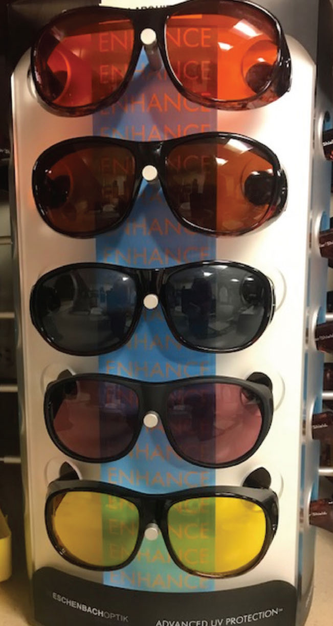
Tinted fit-over glasses for improved contrast and glare management. Click image to enlarge. |
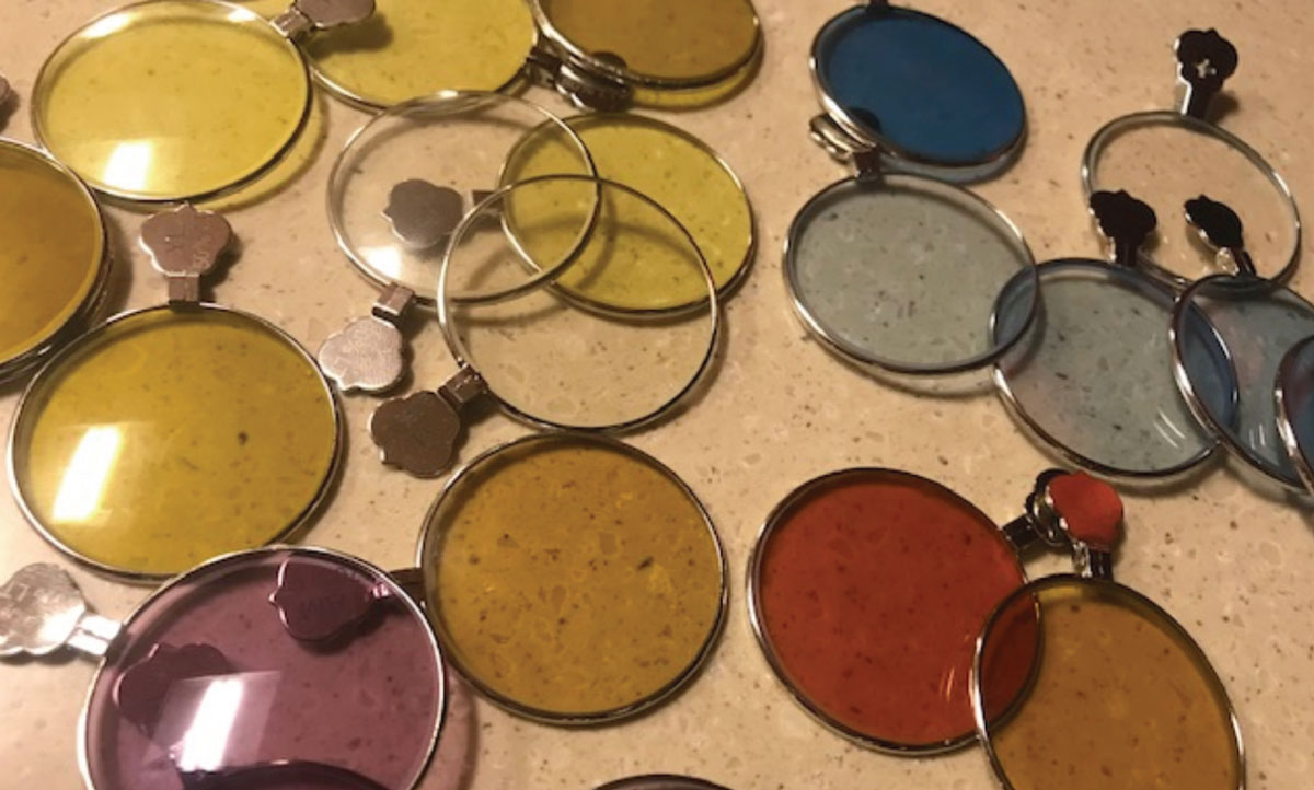
Reduced contrast sensitivity and glare can impact driving by reducing the distinction and clarity of sight in low-light environments or instances where exposure to bright lights is frequent, such as with bright headlights. Additionally, trouble with contrast may hinder mobility by causing difficulty seeing or stepping around objects, while trouble with adaptation to glare may also make independent travel difficult.14 Other tasks such as reading may be impaired as many forms of reading material, such as newspaper, is printed on poorly contrasted material.
To counteract these issues, a thorough tint trial can assist patients immensely. Although the use of tints for LVR is an under-researched area, their use may help improve contrast sensitivity thresholds and cut glare without any impairment of visual acuity.15 If tints are to be recommended for use, LVR specialists will connect with vendors such as Chadwick Optical and Eschenbach Optik to request a tint trial set, which is one of the best ways to complete this evaluation.
Providers should also consider recommendations regarding the patient’s environment. This may include suggesting hats or visors to minimize glare and the amount of light a patient is exposed to. Providing samples of different task lighting may also be useful to enhance contrast. A recommendation may also be made for patients to adopt the use of LED smart bulbs with voice command options. These bulbs often allow for brightness adjustment and can be easy to use for patients once set up in the home.16
Rehabilitation specialists can also be a helpful resource when recommending environmental changes to improve contrast and minimize glare. For example, a lighting evaluation may be completed in the home to provide suggestions to enhance light in certain areas, offer options for better lighting and suggest best locations for reading or completing other visually difficult tasks. Other recommendations can be discussed, such as using dark or light backgrounds on a table to enhance an item’s contrast, making it easier to see.
Diplopia
Occurring when an image is split, usually into two parts, diplopia is generally horizontal, vertical or diagonal. There is a wide range of neurologic causes for diplopia, but common reasons include stroke and muscle palsy.17 Binocular vision disorders such as decompensated phorias may also be a cause.
Diplopia patients may also benefit from a thorough prism trial, as with hemianopsia, which can often be completed in-office. Vision therapy (VT) may be another consideration for patients experiencing diplopia. Like LVR, VT specialists comprise another crucial referral system that primary eyecare providers should be aware of. Provider directory use and reaching out to the nearest optometry school are two possible ways to locate VT providers.
 |
|
Tinted trial rings can show patients different potential tinted lenses for continual wear to enhance contrast and manage glare. Click image to enlarge. |
Takeaways
As most eye doctors do not practice low vision and many areas have limited services, it is imperative that providers at a minimum discuss with their patients their quality of life related to their vision loss. Practitioners should also understand which resources are available in their area to refer patients to. More research is needed to determine which services and practices can benefit patients with low vision, but in the meantime, optometrists must not overlook the importance of connecting the dots between visual function and daily activity impact.
Dr. Capri currently works at Tufts Medical Center, where she specializes in low vision rehabilitation and traumatic brain injury. She also teaches clinical education at the Massachusetts College of Pharmacy and Health Sciences School of Optometry and New England College of Optometry. Additionally, she has an interest in health law and is completing her law degree at Suffolk University Law School. She has no financial interests to disclose.
1. Song Y, Ouchene L, Khan AZ. Saccadic adaptation in the presence of artificial central scotomas. J Vis. 2021;21(1):8. 2. ‘Eccentric viewing’ to improve vision. Lighthouse Guild. Published July 16, 2021. lighthouseguild.org/eccentric-viewing-to-improve-vision. Accessed August 7, 2023. 3. Swienton DJ, Thomas AG. The visual pathway—functional anatomy and pathology. Semin Ultrasound CT MR. 2014;35(5):487-503. 4. Gaffney AJ, Margrain TH, Bunce CV, Binns AM. How effective is eccentric viewing training? A systematic literature review. Ophthalmic Physiol Opt. 2014;34(4):427-37. 5. Cheung SH, Legge GE. Functional and cortical adaptations to central vision loss. Vis Neurosci. 2005;22(2):187-201. 6. Vasconcelos G, Fernandes LC. Low-vision aids. American Academy of Ophthalmology. Published November 4, 2015. aao.org/eye-health/diseases/low-vision-aids. Updated March 4, 2021. 7. Swenor BK, Muñoz B, West SK. Does visual impairment affect mobility over time? The Salisbury Eye Evaluation Study. Invest Ophthalmol Vis Sci. 2013;54(12):7683-90. 8. Chung STL, Legge GE, Cheung S. Letter-recognition and reading speed in peripheral vision benefit from perceptual learning. Vision Res. 2004;44(7):695-709. 9. Moniritilaki M, Badakhsh M, Ehsaei A, Daneshvar R. The effect of contact lens-spectacle reversed Galilean telescope on the visual field of patients with open-angle glaucoma. J Ophthalmic Vis Res. 2020;15(4):502-8. 10. Orientation & mobility (O&M). Commonwealth of Massachusetts. mass.gov/service-details/orientation-mobility-om. Accessed August 7, 2023. 11. Costela FM, Sheldon SS, Walker B, Woods RL. People with hemianopia report difficulty with TV, computer, cinema use, and photography. Optom Vis Sci. 2018;95(5):428-34. 12. Taub MB, Harris P. Fresnel prism to the rescue. Review of Optometry. Published April 15, 2018. reviewofoptometry.com/article/fresnel-prism-to-the-rescue. Accessed August 7, 2023. 13. Peli E. 2017 Charles F. Prentice Award Lecture: peripheral prisms for visual field expansion: a translational journey. Optom Vis Sci. 2020;97(10):833-46. 14. Colenbrander A, Fletcher DC. Contrast sensitivity and visual hallucinations in patients referred to a low vision rehabilitation clinic. Br J Ophthalmol. 2007;91(3):272. 15. de Fez MD, Luque MJ, Viqueira V. Enhancement of contrast sensitivity and losses of chromatic discrimination with tinted lenses. Optom Vis Sci. 2002;79(9):590-7. 16. Lighting and low vision. Perkins School for the Blind. Published December 7, 2022. perkins.org/resource/lighting-and-low-vision. Accessed August 7, 2023. 17. Uzdrowska M, Crossland M, Broniarczyk-Loba A. Is binocular vision worth considering in people with low vision? Klin Oczna. 2014;116(1):49-51. |
