 |
|
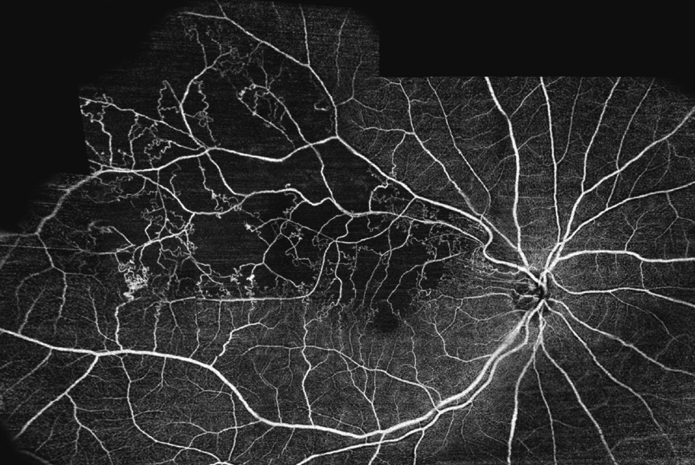
Swept-source OCT-A is a comparable, noninvasive alternative to fluorescein angiography that may save time and improve patient comfort. Photo: Topcon. Click image to enlarge. |
Ultra-widefield fluorescein angiography (UWF-FA) is a valuable test for detecting and monitoring retinal vein occlusion (RVO), but it has several practical drawbacks from cost and time requirements to its invasive nature and tendency to cause nausea, vomiting and even anaphylaxis. It’s also unsuitable for use in pregnant people and those with certain pre-existing conditions such as severe asthma, renal failure and cardiac disease. Widefield swept-source OCT-A (SS-OCT-A), however, avoids some of these limitations. Researchers recently compared the two modalities through a cross-sectional study of patients with RVO lesions, reporting that SS-OCT-A may be a useful alternative modality.
The researchers imaged 34 eyes of 32 patients with treatment-naïve RVO using UWF-FA (200°) and widefield SS-OCT-A. They explained in their paper that the combination of long-wavelength (1060nm) swept-source scanning and a 400kHz A-scan rate is capable of achieving 6mm scan depth and a wide scan area of 24mm x 20mm (81° x 68°) in a single capture, “which may potentially change the paradigm of FA-based diagnosis, grading and follow-up of RVO,” the researchers wrote in their study.
They assessed foveal avascular zone (FAZ) area and perimeter, non-perfusion areas, microaneurysms, capillary changes and collateral vessels.
They reported that FAZ area and perimeter measurements were comparable between the two imaging modalities with high intraclass correlation coefficients suggesting good agreement. They also reported that the mean non-perfusion area was larger on SS-OCT-A, also with a higher intraclass correlation coefficient. Median total microaneurysm count was less on SS-OCT-A than UWF-FA, but agreement in detecting microaneurysms between the two modalities was good. UWF-FA showed fewer total capillary changes and collateral vessels than SS-OCT-A, and agreement between the two modalities was fair.
Overall, the researchers reported that widefield SS-OCT-A was “comparable or even superior in detecting FAZ, non-perfusion area, capillary changes and collateral vessels except microaneurysms in RVO.” Because OCT-A is noninvasive, saves time and is easy to use, the researchers wrote in their paper that this quantitative modality may be a more efficient way to diagnose and monitor RVO.
Li S, Zeng Q, Han X, et al. Comparison of widefield swept-source optical coherence tomography angiography with ultra-widefield fluorescein angiography for the evaluation of lesions in retinal vein occlusion. BMC Ophthalmol. 2022;22(422):1-8. |