 |
|
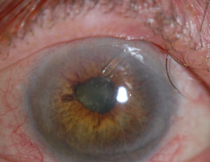
In this analysis, the only significant risk factor for progression was peak IOP after the first three postoperative months. Photo: Peter A. Netland, MD. Click image to enlarge. |
Although trabeculectomy with mitomycin-C has been the mainstay of filtering procedures, there has been a decrease in the number of trabeculectomies performed with a concurrent increase of glaucoma drainage device implantation. Even though IOP is currently the most important known modifiable risk factor for glaucoma, many consider the ultimate goal of glaucoma treatment to be maintaining patients’ visual function and related quality of life. Several studies report visual acuity loss after glaucoma drainage device implantation. Nevertheless, long-term visual field (VF) outcomes have been scarcely described, and more data in this area is needed. Researchers recently measured the magnitude of VF rates of change in glaucoma patients after Ahmed glaucoma valve (New World Medical) implantation and investigated risk factors for VF progression in this setting. They found that there was a continued, significant rate of VF decline after glaucoma valve surgery.
The study, published in the American Journal of Ophthalmology, included 173 eyes. Median IOP and mean number of glaucoma medications were significantly reduced from 23.5mm Hg at baseline to 12.8mm Hg at final follow-up and from 3.3 to 2.2, respectively. VF progression was explored with three methods: mean deviation (MD) rate, glaucoma rate index and pointwise linear regression. Its findings demonstrated that 22% of eyes showed VF progression, and 58% were stable by all three methods, accounting for 80% of all eyes. The rate of VF decline by MD and glaucoma rate index was -0.30dB/year and -2.30 (out of -100), respectively. When comparing progression before and after surgery, the reduction was not statistically significant with any of the methods. The peak IOP (after three postoperative months) was associated with VF deterioration, with a 7% increase in risk per each additional 1mm Hg.
“In our report, 89% of the eyes had a prior ocular surgery, 59% of the patients had prior glaucoma surgery and 27% needed at least one additional glaucoma surgery during follow-up,” the authors wrote in their paper. “It is very likely that our cohort included an important proportion of more complex cases, which might be less prone to achieve VF stabilization after glaucoma drainage device surgery.”
Nevertheless, even though VF progression was not significantly reduced after surgery, the proportion of eyes and rates of progression were fairly acceptable and comparable to other studies. When comparing progression before and after glaucoma drainage device implantation for the subset of 59 eyes with sufficient VFs, the reduction in VF rate of change was not significant.
“While there was a continued rate of postoperative VF decline after surgery, our results compared favorably with rates of VF progression reported by other studies of treated glaucoma,” the authors concluded. “Of all the variables studied, peak IOP after the first three postoperative months was the only significant risk factor for VF progression.”
They also noted that standardization for VF progression assessment would be desirable and then subsequently employed at larger prospective longitudinal studies.
Khaliliyeh D, De Gainza A, Morales E, Caprioli J. Long-term visual field outcomes after Ahmed glaucoma valve implantation. Am J Ophthalmol. March 1, 2023. [Epub ahead of print]. |