 |
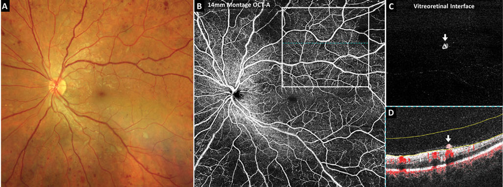
| Using a classification model that included both retinal and choroidal microvasculature helped better identify DR in patients in this study. Photo: Carolyn Majcher, OD. Click image to enlarge. |
Researchers in Singapore have found with swept-source OCT angiography (OCT-A) that flow deficit density with a size threshold better stratified diabetic retinopathy (DR) severity. They have also developed a multivariable prediction model that combined OCT-A metrics from retinal and choroidal microvasculature for DR diagnosis. Their findings demonstrated discrimination between DR and no DR compared with each parameter separately was significantly improved with a classification model that includes retinal and choroidal microvasculature.
The study used 3×3 mm2 fovea-centered scans to evaluate OCT-A parameters from retinal vascular perfusion density, vessel density, fovea avascular zone and choriocapillaris. To compare the discriminative power of these metrics on the presence of type 2 diabetes and DR as well as the need for referral, measurements were compared in three groups. Group 1 compared patients with no diabetes and those with the condition but no DR; group 2 was no DR vs. any DR, and group 3 was nonproliferative DR (NPDR) vs. proliferative DR (PDR).
The study included 35 eyes from 27 unaffected patients and 132 eyes from 75 individuals with diabetes. Severity of DR was classified by no disease (62 eyes), NPDR (51 eyes) and PDR (19 eyes). The data showed that, with the exception of the deep plexus fovea avascular zone area, all retinal vascular, foveal avascular zone and choriocapillaris parameters were strongly altered with DR stages. The researchers also found that stratifying flow deficit density by size and simply calculating it with a size threshold could increase the sensitivity of detecting the rarefaction from both focal and diffuse choriocapillaris degeneration in DR.
Additionally, when compared with retinal parameters, the choriocapillaris parameters allowed the study authors to better distinguish between patients with no diabetes and those with the condition but no evidence of DR.
In their paper on the study, the researchers write that the technique is most effective “in early-stage DR, where the predominant changes happen in the choriocapillaris but not in late-stage DR.” They also speculate that their work might help reveal “differential therapeutic target sites and potential mechanisms depending on the stage of severity.”
Tan B, Lim N-A, Tan R, et al. Combining retinal and choroidal microvascular metrics improves discriminative power for diabetic retinopathy. Br J Ophthalmol. February 9, 2022. [Epub ahead of print]. |