 |
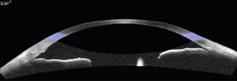
| A recent provided a practical way to evaluate swept source OCT data by focusing on angle parameters measured 750µm from the scleral spur and at the 0° and 270° axes. Photo: Jared Staats, OD. Click image to enlarge. |
Patients who are primary angle closure suspects require greater vigilance to stay out of harm’s way. While anterior chamber depth, an important parameter associated with diagnosis of primary angle closure disease, has been extensively studied, there is limited data on the relationship between it and other anterior chamber parameters in dynamic pupillary conditions. A recent study in Journal of Glaucoma presented key swept-source OCT scan measurements associated with anterior chamber depth change in patients considered to be primary angle closure suspects with cataracts: lens vault and anterior chamber width. The research team believes that examining these parameters may help identify patients at higher risk of developing a severe form of angle closure disease.
The study performed swept-source OCT, at baseline and one hour following pharmacologic dilation, on 78 patients ≥50 years old who are primary angle closure suspects with visually significant cataract. The mean age was 70.9, and 74% of participants were female. Measurements were taken at eight evenly spaced axes at 250µm, 500µm and 750μm from the scleral spur.
Anterior chamber depth change was most correlated with dimension and angle parameters measured 750µm from the scleral spur and at the 0° and 270° axes. The prediction model for anterior chamber depth change after dilation at 0° included: decreased lens vault, wider anterior chamber width and increased trabecular iris space area. The prediction model for anterior chamber depth change at 270° included: decreased lens vault, wider anterior chamber width, larger change in anterior chamber volume, larger baseline anterior chamber volume and smaller baseline angle opening distance.
The researchers noted that the relationship between change in anterior chamber depth and changes in lens vault, anterior chamber width and anterior chamber volume may imply the significant role of lens movement in IOP changes after pharmacological dilation.
While patients who are primary angle closure suspects “comprise the majority of patients having angle closure disease, future studies including eyes with more diverse diagnoses could provide additional insight into the diagnostic power of swept-source OCT in determining risk for dilation-related anterior chamber depth change,” they noted in their paper. “Furthermore, we examined only pharmacologic pupillary dilation and no other dynamic changes, such as in light and dark conditions.”
“By identifying the key swept-source OCT scan parameters associated with anterior chamber depth change in these patients, we provided a practical way to evaluate swept-source OCT data by focusing on these parameters at certain axes, rather than exploring every measurement,” the study stated.
Kao BWH, Yonamine S, Zhao M, et al. Relationship between optical coherence tomography and anterior chamber depth after pupillary dilation in primary angle closure suspects. J Glaucoma. July 22, 2022. [Epub ahead of print]. |