 |
|
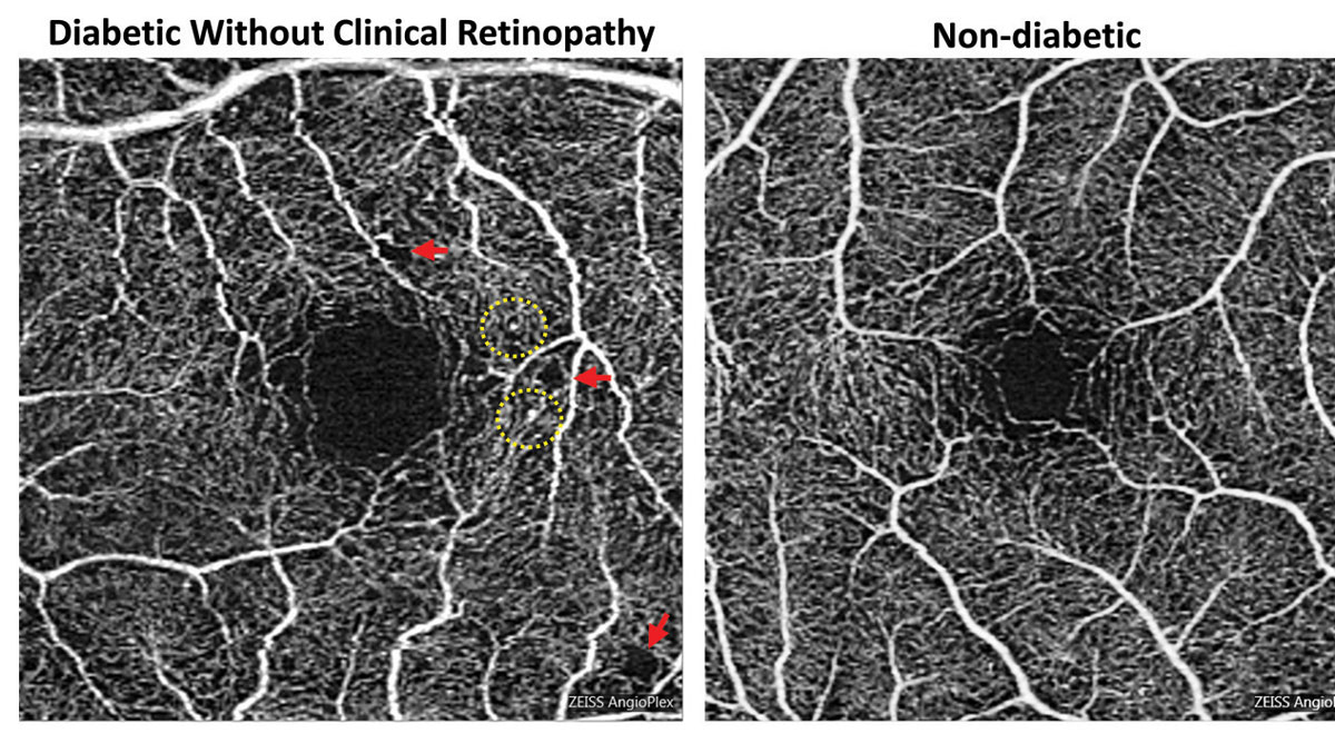
Foveal enlargement and perifoveal capillary remodeling detected with OCT-A in a diabetic eye without funduscopically visible diabetic retinopathy. Red arrows point to subtle areas of capillary nonperfusion, while yellow circles highlight microaneurysms. Photo: Carolyn Majcher, OD, and Susan Ly Johnson, OD. Click image to enlarge. |
OCT angiography can be employed to detect the superficial and deep microvasculature in the retina, which can be useful when assessing diabetic retinopathy (DR). In a recent study, researchers aimed to detect early retinal microcirculation changes in prediabetic patients and investigate their correlation with clinical examination findings.
They reviewed clinical data and OCT parameters of macular vessel diameter (VD), foveal avascular zone (FAZ) and macular vessel area density (VAD) on 147 prediabetic participants: 81 type 2 diabetic patients and 29 controls.
The major findings by OCT-A in the prediabetic group included enlargement and irregularity of the FAZ area, decrease in vessel area density and widening of the mean VD. The pathological change occurred in the superficial and deep layers.
“For instance, in the prediabetes group, VAD in the superficial macular area was decreased and the FAZ size, particularly in the deep layer, was expanded,” the authors explained.
Previous studies reported that the FAZ was enlarged and vessel density was reduced in type 1 and 2 diabetic patients without DR and found evidence of crossing vessels and capillary dropout signs in diabetic patients. That was not the case in this study.
“We did not find the typical crossing vessels sign in the study group, and although some small areas with decreased capillary density were observed, there was no obvious vascular dropout region,” the study authors explained in their paper for the journal Ophthalmic Research. “We speculated that the early pathological changes in prediabetic patients were mild and no typical lesions such as those in patients with diabetic retinopathy were found.”
Capillary changes in superficial and deep layers have been reported by similar studies, but this study could not conclude which layer was supposed to experience a pathological change first detected by OCT-A. “Therefore, a longitudinal cohort study in the future may provide more evidence,” the investigators added. “All these vascular morphological changes were similar to the vasculopathy in early DR but without microaneurysm, which suggested that there may have been lesions in the retinal microvasculature even in the prediabetes stage.”
In the prediabetes group, the axial length was significantly correlated with macular VD and FAZ size, and the urine microalbumin-to-creatinine ratio was correlated with FAZ size. Age had a negative correlation with VAD.
“Our study suggested that enlargement and irregularity of FAZ area, capillary dilation and decrease in vessel area density occur in the retina of prediabetic patients with mild kidney function impairment,” the authors concluded. “Mild increase of urine albumin-to-creatinine ratio could be correlated with early retinal microvasculopathy in prediabetic patients. The results of our study give some hints about the pathomechanism details of the changing microvasculature which guides future OCT-A research.”
Xu Y, Zhu X, Wang Y, et al. Early retinal microvasculopathy in prediabetic patients and correlated factors. Ophthalmic Research. November 16, 2022. [Epub ahead of print]. |