 |
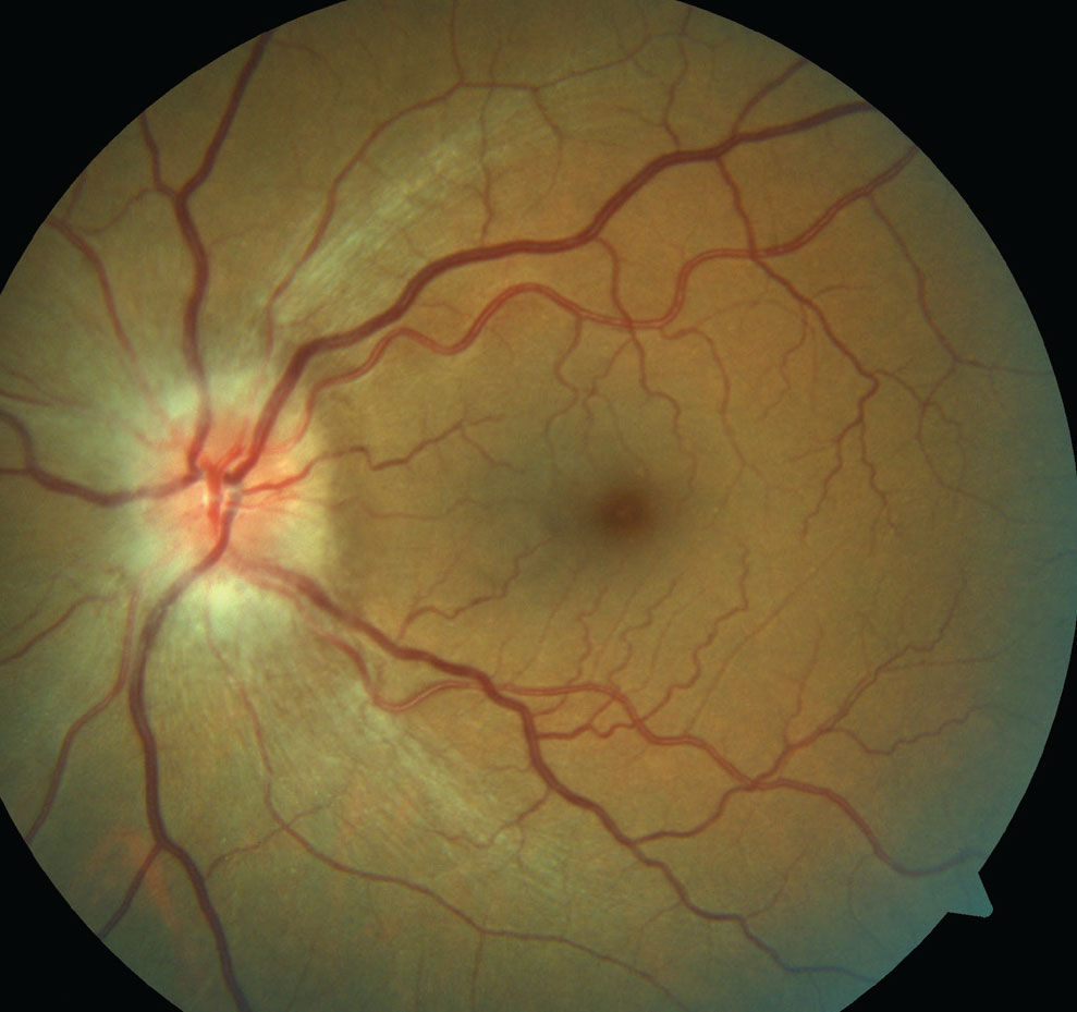
| Minimum rim width may help distinguish between NAION and GON on OCT. Photo: Michael Trottini, OD, and Candice Tolud, OD. Click image to enlarge. |
Clinically differentiating between post-acute non-arteritic ischemic optic neuropathy (NAION) and glaucomatous optic neuropathy (GON) can prove challenging. A recent study, which aimed to identify OCT parameters that may help address this issue, demonstrated that minimum rim width (MRW) is a clinically useful index when distinguishing between these two conditions.
The researchers compared 12 eyes of eight patients with NAION and 12 eyes of 12 patients with GON, which were matched for age and visual field mean deviation. All patients included in this analysis underwent clinical assessment as well as automated perimetry and OCT imaging of the optic nerve head and macula.
The study authors derived the following: neuroretinal MRW, peripapillary retinal nerve fiber layer (RNFL) thickness, central anterior lamina cribrosa depth and macular retinal thickness.
Data showed that MRW was significantly thicker—both globally and in all sectors—among NAION patients when compared with those in the GON cohort. Between the two groups, the difference in MRW increased with increasing visual field loss.
The research team observed no significant difference between groups in RNFL thickness, globally or in any sector. The only exception was the temporal sector, which was thinner in the NAION cohort of patients.
The results also revealed a significantly greater lamina cribrosa depth in patients with GON vs. their NAION counterparts. Additionally, the researchers reported significantly thinner central macular retinal layers in patients with NAION. In terms of the ganglion cell layer, no significant difference was seen between NAION and GON patients.
Based on these findings, MRW was the OCT parameter that most clearly distinguished between GON and NAION, according to the study authors, who noted that this study also suggests that the differences in MRW appear to become more pronounced with increased disease severity.
“The pathogenesis of the increasing MRW difference may be remodeling via the loss of neural tissue and glioarchitecture in GON vs. hypertrophy of the glioarchitecture in NAION,” the researchers concluded in their recent PLoS One paper. “Future studies should longitudinally investigate the unique remodeling of the prelaminar optic nerve head, and the time course of this remodeling, in these two common optic neuropathies.”
Eadie BD, Dyachok OM, Quach JH, et al. Non-arteritic anterior ischemic and glaucomatous optic neuropathy: Implications for neuroretinal rim remodeling with disease severity. PLoS One. May 18, 2023. [Epub ahead of print]. |