 |
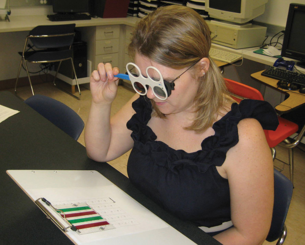
| Type 1 diabetes affects many parameters of accommodation, most commonly accommodative insufficiency. Photo: Deborah Amster, OD. Click image to enlarge. |
Although amplitude of accommodation has been studied in those with type 1 diabetes (T1D), a comprehensive evaluation of all clinical accommodative parameters has not previously been performed. In a new study, researchers assessed accommodative function in non-presbyopic individuals diagnosed with T1D without any signs of retinopathy to determine the existence of possible accommodative disorders related to this disease, as well as the influence of T1D duration and glycosylated hemoglobin values on accommodative function.
The study included a total of 60 participants between 11 and 39 years old, 30 with T1D and 30 controls with no previous eye surgery, ocular disease or medication that could affect the results of the visual examination. Amplitude of accommodation (AA), negative (NRA) and positive relative accommodation, accommodative response (AR) and accommodative facility (AF) were assessed using the tests that showed the highest repeatability. Participants were classified based on normative values into “insufficiency, excess or normal results,” and a diagnosis of accommodative disorders (accommodative insufficiency, accommodative infacility and accommodative excess) was made.
“Subjects with T1D showed a lower ability to accommodate and reduced accommodative facility compared with controls, as well as higher NRA,” the authors explained in their paper. “A higher percentage of accommodative disorders was observed in the T1D group than in the control group, all related to accommodative insufficiency.”
The AA and AF were significantly reduced, while the NRA was higher in the T1D group. More accommodation insufficiency was seen in the T1D participants than in the controls.
“In addition, there was a relationship between the duration of diabetes and AA, AF and NRA, but such a relationship was not seen for the glycosylated hemoglobin values,” the authors noted.
AA was significantly and inversely correlated with age and the duration of diabetes; however, AF and NRA were only correlated with disease duration.
The authors detected accommodative anomalies using the system developed by Scheiman and Wick, which is commonly adopted as a reference for the classification, diagnosis and treatment of accommodative disorders. According to this criterion, those who present with normal values of accommodative function, considering isolated clinical signs, may also show differences in other signs, so they will be added to the group classified with global values of accommodative excess or insufficiency.
“For example, when evaluating AR in the left eye of the T1D group, three, four and 23 exhibited excess, insufficiency and normal values, respectively,” the authors explained. “However, of these 23 subjects with normal values, some also showed differences in other signs and therefore became part of the global group of insufficiency or excess.”
“The main contribution of this study is to note that AA is not the only parameter affected, as both AF and NRA were also altered significantly. Furthermore, from a clinical point of view, the results verify the association of T1D with accommodative insufficiency. AA and monocular AF are clinical diagnostic criteria for accommodative insufficiency.”
The authors added that to their knowledge, this is the only study of similar characteristics where participants were not selected from a hospital but from the general community. The HbA1c value was also determined not only in subjects with diabetes but also in controls.
Silva-Viguera MC, Llamas-Bautista MJ. Accommodative disorders in non-presbyopic subjects with type 1 diabetes without retinopathy: a comparative, cross-sectional study. Ophthalmic Physiol Opt. May 3, 2023. [Epub ahead of print]. |


