Having just experienced a record allergy season throughout much of the United States, and also seeing the introduction of a new topical ocular allergy medication––Lastacaft (alcaftadine 0.25%, Allergan)––we are likely all a bit weary from the recent barrage of allergy education, advertising and “itchy eye” complaints. Many of us were taught to follow the adage, “If it itches, it’s allergy” when addressing the clinical needs of our patients.

And while itching is the hallmark of the allergic response, there certainly are other ocular conditions that can present with varying degrees of itching and similar irritation.
Itching Exposed
Itching, or pruritis as it is called in medical terminology, represents a unique sensory response. A 1964 publication by R.K. Winkelmann, M.D., and Sigfrid A. Muller, M.D., described seven distinct types of itching that may be attributable to a variety of physiologic and pathologic stimuli, systemic disorders (e.g., biliary cirrhosis, diabetes, chronic pyelonephritis and pregnancy) and even psychological conditions.1 The authors further suggested that pain and itching are likely transmitted by the same type of nerve receptors, although they could not pinpoint the exact mechanism which differentiates the two sensations. They provided evidence for several chemical mediators of itching, including histamine, trypsin and bradykinin––all of which may be produced in response to inflammatory bodily reactions.1 Collectively, these chemical mediators are known as pruritogens.
The most recognized pruritogen is histamine. Histamine activates G protein-coupled histamine receptors that are present on a subset of sensory neurons, signaling the brain to perceive an “itchy” sensation.2 Greater concentrations of histamine have been shown to induce a painful sensation, lending credibility to the theory that pain and itch involve a similar pathway.3 But, itching can also be induced by non-histaminic pruritogens; some examples of these mediators include the drug chloroquine and an endogenous substance known as BAM8-22.4
Ocular Itch
Most often, histamine-dependent itch can be managed by histamine receptor antagonists. When dealing with allergic conjunctivitis, relief is usually accomplished with topical agents like olopatadine, epinastine or ketotifen. By contrast, itching associated with non-histaminic pruritogens generally is insensitive to histamine blockers.2 So, although our topical allergy medications are all approved for “itchy eyes,” they may be ineffective when the underlying etiology is something other than allergy. The most common situations in which itching may be reported (aside from allergic conjunctivitis) include anterior blepharitis, meibomian gland dysfunction (MGD), aqueous deficient dry eye, bacterial or viral conjunctivitis, and ocular phthiriasis.
Itching associated with blepharitis may result from a number of scenarios. In anterior blepharitis related to bacterial overgrowth (i.e., Staph. blepharitis), liberated exotoxins set up a mild inflammatory cascade, releasing cytokines into local tissues. These cytokines––including certain interleukins––can, in turn, stimulate pruritus in the skin via specific receptors on sensory nerve cells.5 Likewise, blepharitis can be associated with Demodex, a genus of saprophytic mites that burrow into the base of the hair follicles in humans and animals. Although controversial, species such as D. folliculorum and D. brevis have been identified in patients with chronic blepharitis; their presence seems to be heralded by pathognomonic cylindrical dandruff on the hair shafts that are known clinically as collarettes.6 Itching secondary to Demodex may follow a similar inflammatory cascade as seen in Staph. blepharitis, or the response may be a physical manifestation linked to actual movement of the mites within the skin.
MGD and aqueous-deficient dry eye are most commonly associated with the symptom of burning or grittiness; however, many patients with these conditions also report itching as one of their primary complaints.7,8 The mechanism for this sensation is not entirely clear. Both of these disorders have been suggested to have inflammatory elements, so it is possible that the itching is associated with cytokine production.9 Then again, the itching associated with these conditions simply may represent the low-level firing of pain-sensitive corneal nerves that are stimulated by ocular surface exposure secondary to rapid tear film break-up.

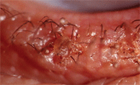
Phthiriasis palpebrarum, as seen here, can cause an itchy, “crawling” sensation.
Infectious conjunctivitis also can be heralded by complaints of ocular itching. Although the typical complaint noted in viral and bacterial ocular surface infections is discharge, patients with mild or moderate presentations also can report burning, stinging or itching.10 By contrast, more severe infections, such as acute bacterial keratoconjunctivitis or epidemic keratoconjunctivitis (EKC), typically present with extreme photophobia and pain.
Finally, phthiriasis palpebrarum represents an infestation of the eyelids and lashes by the genital crab louse, Phthirus pubis.11 One of the principle symptoms associated with phthiriasis is pruritic eyelids. Here again, the mechanism is two-fold. In order to access and feed on human blood, crab lice burrow their heads into the tissue of the lid margins. Similarly to mosquitoes, they secrete saliva, which has anticoagulant properties and can spur an immune response that results in itching, irritation and inflammation of the tissue.12 Also, crab lice respond to various stimuli (e.g., heat, manipulation and chemical assault) by becoming more active, and in doing so may induce a “crawling” sensation that the host perceives as itching.
Differential Considerations
Because numerous conditions can present with ocular itching and other overlapping signs, it is imperative to differentiate these maladies from ocular allergy. One valuable consideration is whether the symptoms improve or worsen when the patient rubs his or her eyes. It is well known that allergic symptoms are exacerbated by digital manipulation; this is presumed to occur because the resultant vasodilation allows increased migration of mast cells and eosinophils to the affected tissues.
Also, mast cells can be manually induced to degranulate with eye rubbing, thus elevating local levels of histamine and other pro-inflammatory mediators.13 Blepharitis, dry eye and infectious conjunctivitis typically improve––at least temporarily––with eye rubbing. Phthiriasis will likely worsen when the lids are rubbed, but direct lid margin observation helps to differentiate this infestation from allergic conjunctivitis.
Careful evaluation allows the clinician to identify blepharitis, although meibomian gland dysfunction (MGD) may not be readily apparent. When considering MGD, you must perform lid margin expression at the slit lamp because many patients with obstructive MGD may not show overt signs of gland inspissation or capping.14 Dry eye has few definitive signs, but in differentiating this condition from allergic conjunctivitis, you should consider fluorescein tear film break-up time and, when possible, tear osmolarity.15
Bacterial conjunctivitis typically is recognized by its associated mucopurulent discharge (particularly upon awakening) as well as pronounced hyperemia, which is stratified toward the inferior aspect of the conjunctiva. Viral conjunctivitis is similar in appearance to allergic conjunctivitis, and can present with comparable chemosis, hyperemia and bilateral involvement. However, the key differential features of viral infection include preauricular lymphadenopathy and a follicular reaction of the inferior palpebral conjunctiva. Also, the RPS Adenodetector (Rapid Pathogen Screening, Inc) provides a swift, inexpensive, in-office test that can identify a viral etiology from the conjunctiva, with a sensitivity of nearly 100%.16
Finally, phthiriasis palpebrarum is often mistaken for acute blepharitis because of its predilection for accumulated lid debris. Eversion of the upper lid helps to expose the burrowing organisms, while recognition of blood-tinged fecal matter and oval-shaped nits on the lashes is diagnostic.
1. Winkelmann RK. Pruritis. Annu Rev Med. 1964;15:53-64.
2. Xiao B, Patapoutian A. Scratching the surface: a role of pain-sensing TRPA1 in itch. Nat Neurosci. 2011 May;14(5):540-2.
3. Broadbent JL. Observations on histamine-induced pruritus and pain. Br J Pharmacol Chemother. 1955 Jun;10(2):183-5.
4. Sikand P, Dong X, Lamotte RH. BAM8-22 peptide produces itch and nociceptive sensations in humans independent of histamine release. J Neurosci. 2011 May 18;31(20):7563-7.
5. Kasraie S, Niebuhr M, Werfel T. Interleukin (IL)-31 induces pro-inflammatory cytokines in human monocytes and macrophages following stimulation with staphylococcal exotoxins. Allergy. 2010 Jun 1;65(6):712-21.
6. Kheirkhah A, Blanco G, Casas V. Fluorescein dye improves microscopic evaluation and counting of Demodex in blepharitis with cylindrical dandruff. Cornea. 2007 Jul;26(6):697-700.
7. Methodologies to diagnose and monitor dry eye disease: report of the Diagnostic Methodology Subcommittee of the International Dry Eye WorkShop (2007). Ocul Surf. 2007 Apr;5(2):108-52.
8. Nichols KK, Foulks GN, Bron AJ, et al. The international workshop on meibomian gland dysfunction: executive summary. Invest Ophthalmol Vis Sci. 2011 Mar 30;52(4):1922-9.
9. Enríquez-de-Salamanca A, Castellanos E, Stern ME, et al. Tear cytokine and chemokine analysis and clinical correlations in evaporative-type dry eye disease. Mol Vis. 2010 May 19;16:862-73.
10. Tarabishy AB, Jeng BH. Bacterial conjunctivitis: a review for internists. Cleve Clin J Med. 2008 Jul;75(7):507-12.
11. Yoon KC, Park HY, Seo MS. Mechanical treatment of phthiriasis palpebrarum. Korean J Ophthalmol. 2003 Jun;17(1):71-3.
12. Jones D. The neglected saliva: medically important toxins in the saliva of human lice. Parasitology. 1998;116 Suppl:S73-81.
13. Greiner JV, Peace DG, Baird RS, Allansmith MR. Effects of eye rubbing on the conjunctiva as a model of ocular inflammation. Am J Ophthalmol. 1985 Jul 15;100(1):45-50.
14. Blackie CA, Korb DR, Knop E, et al. Nonobvious obstructive meibomian gland dysfunction. Cornea. 2010 Dec;29(12):1333-45.
15. Lemp MA, Bron AJ, Baudouin C, et al. Tear osmolarity in the diagnosis and management of dry eye disease. Am J Ophthalmol. 2011 May;151(5):792-8.
16. Barbosa Junior JB, Regatieri CV, de Paiva TM, et al. Adenovirus conjunctivitis diagnosis using RPS Adenodetector. Arq Bras Oftalmol. 2007 May-Jun;70(3):441-4.

