 |
Though Review of Optometry has been the longest-running publication in the field of optometry, this achievement would not have been possible without the continued efforts of our authors—thought leaders who have remained at the forefront of the topics we have covered over many decades. To celebrate them, we asked several for their impressions of articles they authored in a previous era of optometric care to see what they think today of their early work in the field.
July 1960
Tear Flow as a Factor in the Contact Lens Fitting Procedure
By Paul Farkas, OD, and Theodore Kassalow, OD
Today, the buzzword that receives the most attention in the optometric field is “dry eye.” Back in 1960, there were plenty of tears—just ask anyone who went through rigid contact lens adaptation then. Even now, there are still a few older patients who remember receiving their rigid contact lenses and are still happy with them to this day.
Back then, the average contact lens exam room consisted of a projector, phoropter (though a trial frame would do), keratometer, possibly the newly introduced slit lamp, an ultraviolet magnifying lamp and fluorescein. Viewing fluorescein patterns and lens movement were an essential part of the fitting procedure. Many practices also had a hard lens trial set.
 |
For the most part, it was necessary for practices to have on-site lens modification equipment where contact lenses could be modified according to patient symptoms and needs. The addition of edge treatments or peripheral curve modifications could make the difference between success and failure in lens adaptation. Most contact lens manufacturers were initially small mom-and-pop operations that were eventually absorbed into larger organizations. A second important factor was a motivated patient, one who would stick with the optometrist until they were happy or until all other options were exhausted. Bedside manner and handholding were keys to success.
The contact lens material we worked with then was polymethylmethacrylate (PMMA) plastic. We determined the necessary spherical power, center and edge thickness, base curvature diameter and peripheral curves. At the time, PMMA was available in either clear or light tints. Note that making this product work was entirely up to the OD, and though there was some science involved, it was mostly experience and clinical intuition. The cosmetically unacceptable scleral lens was left in the dust in the forties and fifties; by 1960, the “invisible” corneal contact lens was king.
Concerning the tear issue, the challenge first and foremost was the patient achieving lid adaptation to the lens. Frequently noted problems were onset of profuse tearing until the lens settled and then having to live with foreign body sensation until the lid and cornea were desensitized. Though it sounds primitive, keep in mind that these were motivated patients—contact lenses were still a rarity and novelty—who had spent several weeks looking down while being reassured by the optometrist that things would improve. Eventually they did. In fact, with a quality lab and proper edge modifications, a remarkable number of patients did quite well.
The next hurdle was improving wearing time. That meant proper movement of the contact lens to allow tears to circulate freely without slippage, which required modifications of diameter and peripheral curves until the patient goal was met. The final challenge was making certain that the PMMA material remained uncoated during the allotted wearing time. Patients had to receive strict instructions regarding cleaning and avoidance of eye make-up and face creams during lens wear.
To summarize, we did not actually worry about tears too much. Teaching patients to blink properly for optimum tear circulation did the trick. However, the need for the cornea to receive enough oxygen is a story for another time. Today, if an OD saw the level of corneal edema that in 1960 we called central corneal clouding, which many patients lived with, the present-day optometrist would be shocked and demand that the patient instead discontinue contact lenses altogether. But a motivated 1960 contact lens patient would fight against the optometrist for even mentioning discontinuation of that magic piece of plastic that was custom-designed for them. The present-day patient would simply agree with the practitioner and look for an alternate contact lens modality or consider refractive surgery.
Dr. Farkas edits the discussion board ODwire.org, an online community with over 20,000 members. Dr. Kassalow is retired in Sarasota, Fla.
 |
July 1979
Slab-Off Prism Compensation for Anisometropic Vertical Imbalance Induced by Disease
By Jimmy D. Bartlett, OD
This paper—generated from my early interest in ocular disease, nuclear cataracts and other conditions that could potentially cause high anisometropia—was one of my first peer-reviewed articles. The materials for it came from my clinical experiences in the Veterans Health Administration hospital system early in my career. I had just graduated from optometry school, where, like many other optometrists of the time, I was trained principally in vision correction measures, including management of low vision, aniseikonia and other visual anomalies.
For the most part, optometrists then could be described as general all-purpose practitioners, though some more well-known individuals engaged in contact lens specialty practices. Furthermore, optometric curricula in the 1970s was only beginning to include biomedical education as a standard, with a specific focus on biochemistry, human anatomy and physiology, physical diagnosis, systemic and ocular pharmacology and microbiology.
When this article was published, residency education in the mid-1970s was one of the most notable movements occurring in the profession. This optional advanced clinical training has largely been responsible for the recent generation of numerous “subspecialty” clinicians who have expertise in treating ocular disease, pediatric optometry, binocular vision, low vision and specialty contact lenses.
Furthermore, the inclusion of optometrists in the VA hospital program also had a positive effect on the profession’s growth, opening additional channels for practitioners to care for patients with glaucoma, corneal problems and external conditions, in addition to retinal diseases. Overall, the restructuring of the optometric educational curriculum to include biomedical courses as well as the more traditional vision science education along with extensive externship experiences, the inclusion of optometric services in Medicare and many other third-party insurance provider programs, the advent of residency education and access to the Veterans Health Administration system have transformed optometry into a robust primary eye care profession. While challenges continue to exist, these changes over the last several decades have made it so that patients of all ages now receive highly competent and compassionate care from their optometrist.
January 1981
When You Suspect Glaucoma…
By Thomas L. Lewis, OD
The world of glaucoma has changed dramatically in the 35 years since the publishing of this article. Even though the exact cause of primary open-angle glaucoma and the amount of damage it causes to the optic nerve is still unknown, tremendous advances in our understanding of the etiology of the disease at the ultrastructural and biochemical levels still have occurred.
Today, the differential diagnosis of glaucoma also continues to evolve. There is a better understanding of risk factors associated with the disease, which allows for the calculation of the probability of developing glaucoma over a five-year period. This has been accomplished via the discovery of new factors such as the effect of central corneal thickness on IOP readings, as well as a much better understanding of the relative risk of well-known factors including intraocular pressure, race, heredity, optic nerve head appearance and ocular perfusion pressure.
The most obvious impact regarding the diagnosis of glaucoma during this time involves technology. New instrumentation allows us to detect change and monitor over time for both structural and functional damage caused by glaucoma. For example, technology like automated perimetry, confocal laser scanning ophthalmoscopy, pachymetry, more sophisticated tonometry and ocular coherence tomography devices now assist practitioners in refining their diagnosis and treatment efforts. Couple this with normative databases and sophisticated algorithms to interpret the data, and we can not only detect the initial damage from glaucoma, but also determine whether clinically significant progression of this damage is occurring. I am sure that new technologies and a better understanding of this disease will continue to advance the care we provide to glaucoma patients.
April 1983
Senile Macular DegenerationBy Sherry J. Bass, OD, and Jerome Sherman, OD
Upon being asked to update our original article about macular degeneration written in 1983, two facts immediately stood out to us. The first was how long we have both been educators and authors; the second was how much of what we said in this article 33 years ago has changed, while simultaneously still staying part of the scope of the standard of care. So, what are optometrists still doing now, and what has changed?
Back in the early 1980s, the condition mentioned in our original article was known as “senile macular degeneration (SMD)” rather than “age-related macular degeneration (AMD)” as it is called today. The condition’s cause and pathogenesis was also a mystery several decades ago, though it was believed to be the result of sclerosis of the choriocapillaris and/or loss of function of the retinal pigment epithelium (RPE). Research results released then were mixed regarding the relationship between systemic disease and the development of SMD; additionally, no one mentioned the role of green leafy vegetables, and few of us knew about antioxidants.
Nowadays, although we are still theorizing about the condition’s pathogenesis, thanks to ground-breaking research from 2007 we now know that inflammation plays a large role. Other research has also indicated certain dietary trends may also play a role in AMD’s development or lack thereof, including the two AREDS studies in particular, which provide evidence-based suggestions for the use of antioxidant neutraceuticals.
The macular pigments lutein, zeaxanthin and mesozeaxanthin are now significant clinical considerations to monitor for in AMD risk, while tests to measure macular pigment optical density have become useful clinical tools that practitioners can now access. Additionally, genetic testing enables us to provide information to patients regarding their risk for progression based on genetic factors, presence of a history of smoking and body mass index values.
Three aspects of managing senile macular degeneration existed: first, one must find and differentiate SMD; second, one must immediately refer those patients with exudative SMD for treatment; and third, one must provide ongoing specialized care and counseling for patients with this disease. That has not changed.
Binocular indirect ophthalmoscopy was typically performed back then to look for elevation, presence of fluid and other macular abnormalities, and is still done today. Additionally, fundus photography was also performed using a 50-degree fundus camera—still done today—and patients were typically referred to a retinal specialist for fluorescein angiography, which is also still done today. However, nowadays, better technologies for the earlier and perhaps more accurate detection of AMD, the progression of AMD and the development of the exudative changes in AMD now exist. Earlier detection and treatment can help save vision and prevent irreversible damage.
Optical coherence tomography (OCT) is now at our disposal, as well as spectral-domain OCT (SD-OCT) for in-vivo imaging of the retina with excellent resolution that is comparable to histological slides of the retina. OCT imaging can help practitioners identify any present drusen, pigment epithelial detachment or serous detachment, which may be due to the presence of choroidal neovascularization (CNV). Interestingly, while standard SD-OCT may not pick up choroidal neovascular membranes in exudative AMD, recently, a newer type of OCT known as OCT angiography (OCT-A) has also become available. This technology can detect possible CNV membranes without the use of dye injection.
Several decades ago, the only way to monitor the progression of atrophic AMD was to perform direct and binocular indirect ophthalmoscopy, as well as fundus photography. In 2016, however, OCT is now being used to detect the progression of atrophy of the outer retinal elements. Fundus autofluorescence is also in use to detect changes that are not evident even with color photography or ophthalmoscopy. These technologies will likely play an important role as drugs that slow or even prevent the progression of dry, atrophic changes continue to arrive on the market.
Regarding the referral system back then, most practitioners would refer their patients to retinal specialists for the treatment of exudative AMD. This is still the case now; however, the only treatments available then were blue-green laser and red krypton laser for the treatment of juxtafoveal and extrafoveal CNV. There was no treatment for subfoveal CNV back then, and thus we had to watch many of our patients’ vision deteriorate as the CNV turned into a massive disciform scar. So many lives were affected then. Now, however, intravitreal injection of anti-VEGF agents has changed the destructive course of the wet form of this disease in many patients. Though a chronic and essentially lifelong injection regimen is still currently required to control the condition, the future holds promise for better injection schedules as even newer agents become available.
September 1985
Easing into Automated PerimetryBy Paul C. Ajamian, OD
It is so interesting to think of automated perimetry, considered so routine today, as revolutionary—but at the time, it was. In the 1970s and early 1980s, manual Goldmann kinetic perimetry was the standard. It took a lot of time, experience and active involvement from the operator and created angst in many patients as well. The early Omni comanagement center directors all paid their dues by doing many 5 isopter Goldmann fields!
 |
The 1985 article covers several topics, including supra-threshold screening perimetry in comparison with threshold tests. I still believe in screening perimetry, though it is rarely used. If you are testing for retinitis pigmentosa, headaches or a hemianopic defect from a stroke, there is nothing better and faster than a supra-threshold screening field such as an 81 or 120 full field. However, most offices are in “threshold mode” and order a 24- or 30-degree threshold field for everyone and everything—not always necessary, for sure.
The article also mentions that it typically took 12 to 18 minutes per eye to perform 30-degree testing—something which newer technology and ongoing research has altered to 24-degree testing performed in just a matter of minutes. That duration of testing wouldn’t fly in today’s busy office, nor would patients tolerate it well. Additionally, the early automated units cost $10,000 or less, compared to today’s price tag of well over $20,000, even with a trade-in.
It is fun to look back knowing that our early automated perimeters seemed like something out of a science fiction movie, but they served as the foundation upon which today’s modern instruments were built.
February 1986
Targeting Extended Wear Trouble Spots
By Joel A. Silbert, OD, and Frank D. Fontana, OD
Joel: It was an honor to have been included in the forum 30 years ago on extended wear led by “Uncle” Frank Fontana, as well as the late Roger Kame, Rex Ghormley and Jack Solomon, and two young lads (Ken Lebow and yours truly). It was also interesting to read the transcript of that session in which both hydrogel extended wear and the newer modality of gas permeable extended wear was discussed. As promising as GP extended wear was—in part based on its higher safety profile—it never really took off, mostly due to discomfort issues associated with GP lens wear. The irony today is that GP extended wear is routinely used in orthokeratology treatments and is highly successful.
Thirty years ago, traditional orthokeratology was unpredictable and not highly effective: it took the development of high Dk polymers, reverse curve technology and corneal topography to make it the highly successful mainstream option that we enjoy today. Much of the discussion during that forum revolved around concerns of complications from hydrogel extended wear and patient noncompliance. There were indeed many complications that resulted from 30-day extended wear with hydrogel lenses, including many cases of corneal edema from snugly fitting lenses. Today, in contrast, we can look back and take comfort in knowing that polymer science has essentially eliminated certain complications of extended wear. For this, we owe gratitude to the science involved in the Nicolson patent that led to the development of the silicone hydrogels that many of us take for granted today.
Despite the advent of these products, many modern practitioners still face challenges with silicone hydrogel extended wear that are not specifically edema-driven, but rather are caused by ocular surface issues and/or bacterial adherence. These include contact lens papillary conjunctivitis (CLPC), infiltrative keratitis, contact lens-induced acute red eye response (CLARE) and microbial keratitis. Although the severity of these complications is tempered by silicone hydrogel lenses, they still exist. Furthermore, patient compliance issues still exist and, as such, many of us see benefits stemming from daily disposable lens wear that were not in existence 30 years ago. Many patients enjoy the convenience and good eye health associated with these products, which are now in ready supply. Daily lenses have also led to greater use of contact lenses by children and teens, helping to allay fears of noncompliance by these groups of patients. Thus, there is less need for extended wear.
Frank: In those days, we had to be careful of who we chose as a patient. You couldn’t be too sure whether a patient would follow your instructions. With the commonality of two-week overnight wear, any noncompliance on the part of the patient could lead to serious ocular complications at a time when medications to treat these problems were still under development. Lens materials and lens care solutions have also improved significantly in the last several decades, as have lens options.
The best thing that ever happened in the industry was the introduction of daily disposable lenses, as they have increased patient compliance with keeping lenses clean—and eyes healthy. The only downside is that daily disposable lenses cost more than their earlier counterparts. Still, the benefits outweigh the negatives.
 |
February 1987
How to Fit Rigid Gas Permeable Extended Wear Lenses
By Joseph P. Shovlin, OD
As I look back on the last three decades and reflect upon contact lens technology and lens options in particular, I am amazed at just how successful we were back then. So, the question is, were we luckier than we deserved to be, or were we forced to pay particular attention to the things we really understood at the time?
Back then, topography/tomography systems did not exist, and neither did the modern point-of-care diagnostic technologies available today. Lens options and solutions were also significantly more limited; yet at the time, we fit both GP and hydrogel extended wear lenses with some impunity from the dreaded complications with which patients were adequately screened and educated upon.
Nowadays, things have been tempered a bit. Extended or continuous wear is rarely used as a viable lens option for new patients today, which may, in part, be due to additional concerns that we have learned over the years, though it could also be the result of expanded technologies like daily disposables and refractive surgery options. I hope that new advances and additional options will continue to bolster the profession.
May 1987
Glaucoma: How to Evaluate the Visual Field
By Murray Fingeret, OD
Wow, how time flies. Amazingly, the principles in the article (especially the steps in analyzing the field) are still applicable, though some things have changed since 1987. These changes include the introduction of Statpac Total & Pattern deviation maps, which revolutionized the analysis of single fields and was further refined with the arrival of the glaucoma hemifield test. Furthermore, glaucoma change probability analysis introduced the concept of event analysis for change, later strengthened by the arrival of the early manifest glaucoma criteria for interpreting it.
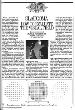 |
Additionally, short wavelength automated perimetry came and went in popularity, while the concept of requiring a visual field defect to define someone as having glaucoma is also no longer pertinent in today’s modern practice. Instead, the presence of optic nerve damage or retinal nerve fiber layer loss is now consistent with a diagnosis of glaucoma, even in the case of a full visual field. Lastly, the advent of HFA2 technology meant better patient ergonomics and gaze tracking.
Our attitude about the significance of detectable glaucoma change has also morphed radically. Before 1996, even the tiniest detected change demanded an escalation in therapy, no matter how radical. After 1996, thought leaders began basing therapeutic decisions on how fast change was happening, life expectancy and stage of the disease—not just the detection of any amount of change. It took another 10 years for this idea to permeate eye care, but it started in 1996. This new idea was introduced by Michael Patella, an optometrist who works at Carl Zeiss Meditec and was the developer of the HFA perimeter.
Very little has changed in the last 12 years, but a lot changed between 1987 and 2003, and more changes are on the horizon. Advances in the near future include tests to allow optometrists to examine the macula in more detail, tests that are expected to take half the time of the SITA Standard and the introduction of Size V testing, which will help reduce the variability and refractive error effects and also extend the test range.
May 1989
Sharpen Your Retinal Exam Skills
By Leo P. Semes, OD
A quarter century ago at the time of this article, optometry had embraced dilated fundus evaluation. The year 1989 also saw Kareem retire, Pete banned from baseball, Tiananmen Square shake the world, the Exxon Valdez run aground and Time select Gorbachev as man of the year (as opposed to person). Just a year prior, I, together with my colleagues, John Potter, Tony Cavallerano and Matt Garston, published our primer on performing binocular indirect ophthalmoscopy (BIO) and scleral indentation. At the time, autorefractors were primitive at best, online contact lens and spectacle sales were relatively unknown and eye examinations were performed live.
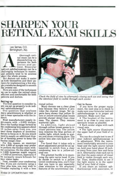 |
Contemporarily, the profession enjoyed nationwide dilation privileges. Fundus evaluation has largely evolved to survive along with BIO and posterior pole scrutiny with high dioptric-power plus lenses (+60, +78). These lenses offer enhanced stereopsis over a +90D. Yellow lenses have not been widely available, but with the current interest in phototoxicity, they may yet make a comeback. Pupillary dilation is now more convenient for both patient and clinician with the availability of Paremyd (0.25% tropicamide, 1.0% hydroxyamphetamine, Akorn). Though this formulation was available in 1989, it was not widely used and became dormant. However, now, I find that this formulation—available again—works very well for most patients, though it may be less effective in the elderly. It was conceived by an optometrist, the late Dr. Michael Larkin.
Also today, scleral indentation is taught routinely in optometry schools and is used widely by optometrists. BIO instruments have also become lighter and the light sources have evolved to LED, which offer better illumination and, consequently, improved resolution.
Interestingly, the patient discussed in the original article is now a practicing optometrist in Hayes, Va. Dr. Peter Wilcox states, “Today’s technologies have moved many fundus structures and conditions from the clinically occult to the appreciable and hence, actionable, for the diagnosis, treatment and management of disease. For instance, the SD-OCT machines and certain angio-scanning devices (OCTA) allow for a sometimes-decades-earlier detection of posterior segment disorders. This allows patients to be counseled sooner to modify or initiate changes for more positive outcomes. The scanning laser ophthalmoscopy (SLO) technologies also allow for the never-dreamed-of-before-1989 analysis of ocular structures, and the upcoming shift in classic visual field analysis from gray tone printouts of visual field losses to the direct overlay (objective perimetry) of deficit to the deficient ocular structure or the pipeline of the neuro-ocular disease may also benefit practitioners.”
It’s clear that OCT technology as well as certain other structural imaging devices now have penetration among optometrists comparable to what BIO had in 1989. Looking forward, we will likely see greater use of auxiliary evaluation strategies such as fundus autofluorescence, automated dark adaptometry, ultra-widefield fundus imaging as well refinements in the hardware and software for OCT. These devices not only augment our clinical observations but also help us better understand ocular and systemic disease. We’ve come a long way in retinal examination in a generation.
September 1989
Is Optometry Ready for Laser Surgery?
By J. James Thimons, OD
Atomic physicist Dr. Niels Bohr once said, “It is exceedingly difficult to make predictions, especially about the future.” This article on laser surgery published in Review of Optometry 25 years ago makes reasonable truth of his off-handed observation on quantum physics. As it turns out, the nature of our profession and its progression during the last few decades has been just as difficult to predict.
Who would have thought over 25 years ago that optometrists today would have glaucoma privileges granted in nearly all 50 states (Massachusetts being the lone holdout), and that we would also be accepted as clinical partners in various national healthcare systems, Veterans Administration Medical Centers and large HMOs, both regional and national? The advent of laser surgery proved itself to be akin to Dickens’ A Tale of Two Cities, as the industry burgeoned mightily and almost succeeded in manifesting the predictions of some of my good friends and colleagues, who viewed it as the start of our profession’s demise. Instead, while still present, laser surgery is now a notably declined market, with no achievement of our long-term fears of world dominance.
“Within 10 years, lasers will be an integral part of patient management. It’s almost impossible for optometry not to be involved,” I wrote back then. I was prescient enough to contextually discern this prediction at the time, but not appropriately define. For more than a decade, Northeastern College of Optometry has been training young, vibrant and talented individuals to provide laser vision correction for their patients at both the college and in their practices following graduation. This is a program that didn’t seem possible to run back in 1989.
Furthermore, I don’t think that any of us would have predicted the development of optometry outside the United States 25 years ago. Since then, however, it has been a delight to work together with provinces and states in both Canada and Australia, especially as they continued to advance their level of clinical practice. Most recently, I was honored to witness and participate in the nationwide, singular legislative enactment that provided authorization for optometrists to treat glaucoma in Australia. This remarkable accomplishment has remained relatively under-appreciated here in the States given the distance between the countries and somewhat remote connection that many of us have to that part of the world. However, it is one of the biggest accomplishments that we have had in our profession, in that we have begun the process of universalizing the profession that we all love so very much.
As we did at the onset of laser technology, I believe it is our responsibility to continue adapting to and capitalizing upon other new innovations still in the works, from optical systems to disruptive technologies involving online access and diagnostic and therapeutic regimens, both medical and machine. In many instances, they are either at the margin of or beyond the current reach of our legislative scope—but this should only invigorate us to work more diligently to achieve better coverage, keeping in mind that they will be to the future what laser vision correction has been to us so far.
It has been a wonderful opportunity to share over 25 years with Review of Optometry as a colleague, contributor, reader and fan. I hope for the magazine and for all of us that the next 25 years will be as exciting and as fruitful as the last have been, and that the future will also continue to hold both the enjoyment and also the mystery of Dr. Bohr’s insightful perspective.
 |
September 1989
An Inside Look at the New ‘Secret’ Agents
By Christopher J. Cakanac, OD
I wish I still had the hair I did when I wrote this article in 1989. Madonna was on the radio, and I was wearing skinny ties. At the same time, a pharmaceutical rep visiting an optometric office was such a rare occurrence that this article was primarily an attempt to get the message out about new drugs. Though many of those drugs are now gone, this article did touch upon the infancy of two drug classes we still use extensively today: topical fluoroquinolones and nonsteroidal anti-inflammatory agents.
To appropriately treat corneal ulcers in the 1980s, practitioners required adequate access to fortified topical antibiotics, the standard of care of the era. They had to be custom compounded, usually in a hospital pharmacy, which, for the most part, prevented optometrists from using them routinely. Norfloxacin was the first commercially available fluoroquinolone; other drugs including Ciloxan, Vigamox, Zymaxid and Moxeza also eventually gave optometrists greater ease of treatment for corneal ulcers with readily available agents.
Another drug, Ocufen, was the first of a separate new class of medications known as nonsteroidal anti-inflammatory agents (or NSAIDs). Other more recent examples include Voltaren, Acular and Prolensa. Drugs with anti-inflammatory activity that didn’t have the side effects of steroids were unheard of at the time. Ocufen was the first NSAID introduced, and its indication was to prevent miosis during cataract surgery, though we quickly learned it prevented cystoid macular edema, which is still one of the primary indications for NSAID use today. We also discovered these medications prevented pain from corneal abrasions, thus eliminating the dreaded pressure patch.
May 1996
The Clinical Guide to Ophthalmic Drugs
By Ronald N. Melton, OD, and Randall K. Thomas, OD
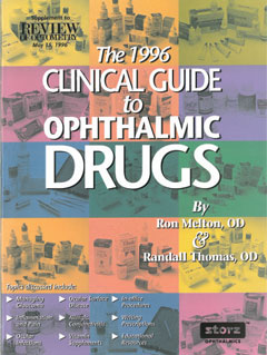 |
When we started optometry school back in the fall of 1977, North Carolina had become only the second state to authorize legislation for the prescribing of therapeutic drugs by optometrists. We were keenly aware of this new high bar that was set for our profession, and for the next four years that we attended the Pennsylvania College of Optometry, we dedicated ourselves to learning all we could about therapeutic eye care. During that time, it became clear to us that there was a gap in pharmaceutical/therapeutic education in the post-doctoral optometric population. As such, we felt the need to develop some kind of a concise, clinically practical guide to help speed the acquisition of knowledge in the field of ocular therapeutics. This effort began about 20 years ago, when we sat down to put together the first drug guide.
Over the years, many medications have come to market beyond what was initially offered to the first optometrists to gain TPA rights and, beyond that, there has also been an expansion in the use of currently available drugs. Our purpose with the drug guide was not only to educate our colleagues about new drugs but also to enhance our colleagues’ therapeutic application of those drugs that were currently available at the time. More than anything, we wanted to instill greater confidence in our colleagues to embrace this vital aspect of optometric practice.
It is in this spirit that we continue to edit the publication on an annual basis, now in its 20th year. Our work with the drug guide has, in a large part, been motivated by a great many of our colleagues who have encouraged us to continue in our efforts. We sincerely wish there were even more new drugs and applications to discuss annually. Fortunately, the professional literature continues to provide us with additional information to share. It is our hope that the drug guide will continue to be a help to those in our profession who strive to achieve a greater level of excellence in patient care.