Anatomical structure studies stood out as a theme at ARVO 2013, perhaps a consequence of glaucoma investigators now taking full advantage of optical coherence tomography innovations that allow for unprecedented visualization and data acquisition of the posterior segment. One can only hope a reliable structural metric for diagnosing and/or measuring glaucoma progression will soon emerge from these nascent efforts.
In the meantime we still have visual fields as a mainstay, which researchers continue to improve upon. Blood flow—sometimes known as intraocular pressure’s dark twin—remains largely mysterious, but gave up a few secrets this year. Attendees also perused data on a new class of glaucoma drug, plus an interesting punctal plug implanted in dogs.
Structural Metrics
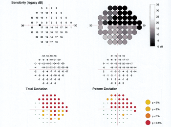
Using enhanced-depth imaging spectral-domain optical coherence tomography (EDI SD-OCT) and the Humphrey visual field test, researchers at the University of Washington School of Medicine in Seattle found a correlation between lamina cribrosa (LC) morphology and glaucoma severity.2253/B0057
Optic nerve head B-scans of 103 glaucoma patients were obtained using EDI SD-OCT. Images were analyzed using the Heidelberg Eye Explorer software. LC depth was defined as the greatest distance between the reference plane (the imaginary extension of Bruch’s membrane plane) and the anterior border of the LC, perpendicular to the reference plane. LC thickness was the distance from anterior border to the posterior border of the LC in the center of optic nerve head images.
In this study, researchers found that glaucoma severity shows a significant correlation with LC depth and LC thickness, metrics that may be useful during optic nerve head analysis.
Peripapillary choroidal volume (PCV) appears to be reduced in eyes with glaucoma compared to healthy eyes or ones with ocular hypertension, according to a paper presented by the University of Houston College of Optometry.2150 A total of 123 subjects participating in a longitudinal, observational clinical research study and diagnosed with primary open-angle glaucoma, ocular hypertension or deemed normal were imaged using SD-OCT, again with enhanced-depth imaging. Whether thinner PCV reflects an inherent risk of POAG or occurs during glaucoma’s pathogenesis requires further study.
A new OCT technology that visualizes macular RNFL loss at different stages of glaucoma appears to correlate well with visual fields, according to research teams in Milano and Bergamo, Italy.4815/D0254 Ten healthy eyes and 10 eyes with varying stages of glaucoma underwent RNFL transversal scans using the incorporated analysis software of the Heidelberg Eye Explorer (version 5.7.0.1).
The new scan’s dense volume allows for detection of RNFL loss, from early to advanced stages of glaucoma. This visualization follows the same visual field damage shape as anatomical RNFL distribution. The OCT’s quicker speed could make this new approach useful in clinical practice, investigators believe.
Another novel OCT measurement, macular retinal ganglion cell analysis via both the Cirrus HD-OCT and RTVue-OCT, appears to work well, according to a Parisian study.4830/D0269 A total of 167 eyes were split into early glaucoma, moderate-to-advanced glaucoma and healthy groups. The two ganglion cell analyses correlated well with circumpapillary RNFL measurements from each machine respectively, plus both machines showed a similarly high sensitivity and specificity, researchers concluded.
Yet another biological marker—the inner-to-outer retina area ratio—appears to also hold promise as a glaucoma detector, according to a study team at the Jules Stein Eye Institute.4840/D0279 Forty-one normal subjects and 27 glaucoma patients underwent study. When compared to ganglion cell/inner plexiform layer thickness measurements, the inner-to-outer retina area ratio was found to be significantly predictive of glaucoma. The clinical benefit is that this new measurement does not vary as a function of age or axial length in normal subjects, the study found.
Researchers in Hiroshima attempted to determine how glaucomatous damage affects the thickness of three important structures: retinal nerve fiber layer (NFL); the ganglion cell layer and the inner plexiform layer (GCL+IPL); and the outer retinal layer (ORL).4851/D0290 Eighty-four glaucoma patients and 36 normal control subjects were studied using OCT. The NFL and GCL+IPL measures in the normal group were significantly thicker than those in the glaucoma group. The outer retinal layer in glaucoma patients was thinner than that in normal subjects, but not to the point of statistical significance. Researchers concluded glaucoma mainly damages the inner retinal layer in the macular area, but that loss of the outer retinal layer may occur too. Also, they suggested that damage to the NFL precedes damage to the GCL+IPL.
Structure vs. Function
We always hear about structural and functional changes that occur as glaucoma progresses. But what about as treatment progresses, when IOP is reduced? A team at Wills Eye Institute looked into this question.1877/B0131 Forty-seven glaucoma patients with proven pressure-lowering interventions were studied.
Change in IOP was significantly associated with change in cup volume and rim area after the first follow-up visit. Change in functional vision approached significance after the first follow-up visit in patients with the most drastic pressure reduction, but fell just short.
Change in RNFL thickness over time also fell just short of significance among drastic pressure-reduction patients as well. Thus, marked reduction of IOP in glaucomatous eyes caused some early structural changes but no significant changes in functional tests, researchers concluded.
Researchers from Sao Paulo sought to evaluate the correlation between peripapillary retinal nerve fiber layer thickness and visual field indices using different SD-OCTs and visual field perimeters in glaucoma.2283/B0087 In a total of 44 eyes of 25 patients included in the study, moderate correlations between peripapillary RNFL thickness and visual field indices were found, according to the paper.
A big question in all structure vs. function discussions—which sustains damage first?—was addressed in an interesting way by a multi-site US study team.4939 They investigated the tendency for the conservation of the binocular visual field in patients with moderate to severe glaucomatous field loss in both eyes.
The researchers studied 47 patients with stabilized chronic progressive glaucoma undergoing Humphrey Visual Field 30-2 testing and found a very strong tendency for optimizing the binocular visual field in a manner that defied simple anatomic symmetry considerations. The paper noted that focal axonal injury in one eye may be accompanied by increased activity in the contralateral retinal glia and geniculate layers receiving visual information from the fellow, non-injured eye. Focal bilateral compensation of this kind, mediated by the body’s central nervous system, may be involved in the conservation of the binocular visual field in patients with glaucoma, the study observed.
Visual Fields
Optical coherence tomography is a wonderful resource, to be sure, but there is still a great deal of diagnostic value to be had from visual field (VF) testing as well. An international study group made an enhancement to its visual field risk calculator.2631 Despite good performance in predicting final mean deviation, the calculator failed to account for the localized nature of glaucomatous disease and progression, so it could not predict sectorial VF deterioration. Researchers aimed to enhance it by adding visual field topographical information, validating the improvement in a subset of 367 glaucoma patients. The enhanced calculator proved accurate at estimating glaucomatous progression in different sectors, which may be more helpful than global indices for assessing areas of greater risk of progression and estimating the rate of progression in each VF sector, the study suggested.
A US study team found that personalized examination schedules may improve the likelihood of detecting glaucoma progression.3956/D0244 A total of 571 glaucoma patients underwent evaluation, some receiving a visual field examination and IOP check once a year on a fixed schedule, and some who were examined at intervals based on what the researchers called the Kalman filter, which was validated by data from the Collaborative Initial Glaucoma Treatment Study (CIGTS) and the Advanced Glaucoma Intervention Study (AGIS).
Flicker-defined form perimetry, as demonstrated here on the Heidelberg Edge
Perimeter, yields fewer fixation losses and a lower rate of false positives than
standard automated perimetry testing. Photo: James L. Fanelli, OD
The model forecasts each patient’s disease dynamics into the future while incorporating the uncertainty associated with those forecasts. Researchers showed a 27% increase in efficiency in detecting progression among those on personalized schedules.
Flicker-defined form (FDF) perimetry was designed to detect early VF loss in glaucoma. Researchers at Moorfields Eye Hospital in London sought to compare this test with standard automated perimetry (SAP) using a cohort of 137 patients.3952/D0240 They found the FDF test to be associated with higher false negatives than SAP. However, patients performed better on the FDF in terms of fewer fixation losses and lower false positive rates. Additionally, the FDF test duration was faster, researchers concluded.
Frequency doubling technology (FDT) perimetry can be used to track glaucoma progression, according to a study based in Groningen, Netherlands.3927/D0225 Among 126 glaucoma patients who were followed for about six years with both the Humphrey Field Analyzer (30-2 SITA) field testing and FDT (C20-1 full threshold), there was a highly significant correlation of results. This suggests that FDT can be used in patients who cannot be reliably tested with SAP.
SLT Lasers
Selective laser trabeculoplasty (SLT) is effective at reducing IOP in both initial and repeat treatments in patients with pseudoexfoliative glaucoma, according to a two-team study based in Massachusetts.1857/B0111 Investigators studied 56 patients and a total of 79 eyes that underwent as many as three SLT treatments over 11 years.
The percentage of eyes maintaining IOP control without additional SLT (about 50%) and those requiring surgery (about 20%) were similar after the first and second SLT treatments. Although a small group (three of five patients), 60% of eyes required surgery after the third SLT treatment, the study found.
In patients receiving maximal medicinal treatment, SLT as a secondary treatment showed efficacy in reducing or maintaining IOP for three years, according to a study conducted at Wills Eye Institute.1866/B0120 Eighty-eight glaucoma patients (75 with POAG, six with secondary glaucoma, and seven with normal-pressure glaucoma) who received three or four medicinal treatments were followed up after treatment with SLT at one-, three- and five-year intervals. Mean IOP was calculated before receiving laser treatments then compared to the IOP measured at those same intervals. The mean baseline IOP was 18.35mm Hg. The mean at three years was 15.86mm Hg.
Blood Flow
For years it has been postulated that problems with blood flow to the optic nerve head may explain the phenomenon of normal-tension glaucoma. At the heart of these theories is a process called autoregulation. This prompts the question: are certain categories of patients prone to autoregulation dysfunction?
An international study team tackled this question for diabetics.4472/D0212 They examined the relationship between systemic blood pressure (BP) and ocular perfusion pressures (OPP) with blood flow in the temporal (TPCA) and nasal (NPCA) short posterior ciliary arteries in 75 open-angle glaucoma patients, some with and some without diabetes mellitus.
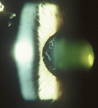
Selective laser trabeculoplasty is effective at lowering IOP in patients with pseudoexfoliative glaucoma, as seen here. Photo: Paul C. Ajamian, OD
In diabetics, changes in short posterior ciliary artery blood flow were strongly correlated to changes in BP and OPP. These correlations were weak among those without diabetes; the differences were statistically significant. This data suggests that diabetic glaucoma patients may have impaired vascular autoregulation during fluctuations in systemic BP and OPP, according to the study.
An Indiana University School of Medicine study looked at this question among African Americans.4470/D0210 The researchers examined differences in the relationship between systemic BP and OPP with localized ocular blood flow in the central retinal artery (CRA) and ophthalmic artery (OA) in 56 glaucoma patients of European descent (ED) and 19 of African descent (AD).
In glaucoma patients of AD, CRA blood flow was more strongly correlated to blood pressure and perfusion pressure than in patients of ED. OA changes were more strongly correlated to ED than AD patients. This indicates that retinal blood flow may not be sufficiently autoregulated during changing blood and perfusion pressure in patients of African descent, according to the study.
Novel Topics
It would not be ARVO without a look at potential new breakthroughs. Here are but a few.
An industry-sponsored US study team investigated the efficacy and tolerability of trabodenoson, a highly-selective adenosine-1 agonist, which is a new class of glaucoma drug.2621 One hundred and forty-four subjects were randomized to placebo or trabodenoson. The most consistent decreases in IOP were noted at the 500μg dose, the study found, concluding that the drug was well-tolerated, safe and resulted in significant IOP reductions in adults with ocular hypertension or POAG.
Endoscopic cyclophotocoagulation (ECP), a new IOP-lowering procedure that can be combined with cataract surgery, is a safe and simple strategy for the long-term control of mild to moderate glaucoma, a multi-site US study team concluded.4750/D0134
The mean baseline IOP in 261 eyes of 163 patients was 17.27mm Hg, which was significantly reduced at every time point from baseline to 66 months, with average IOP at month 66 being 13.63 mmHg among patients who had undergone ECP. That amounts to an average IOP reduction of 21% over 66 months. After ECP, patients on topical medication were 10.5 times more likely to be off of medications at 66 months when compared to baseline, the study found.
It appears punctal plugs made from the drug travoprost can effectively serve as a sustained release modality, at least in canine models thus far, according to an industry-sponsored US study.5633/C0019 One-, two- and three-month sustained release of travoprost from biodegradable punctal plugs is feasible, the study concluded, and can deliver travoprost into the tear fluid at therapeutic levels. Plug retention is a key attribute to achieve clinical success, and confirmation of plug presence can be performed by physicians or patients, the study noted. Researchers confirmed visualization and retention of the canine plugs.
